Embryo series courtesy of Einhard Schierenberg
Table of Contents
Abstract
Transposons are discrete segments of DNA capable of moving through the genome of their host via an RNA intermediate in the case of class I retrotransposon or via a "cut-and-paste" mechanism for class II DNA transposons. Since transposons take advantage of their host's cellular machinery to proliferate in the genome and enter new hosts, transposable elements can be viewed as parasitic or "selfish DNA". However, transposons may have been beneficial for their hosts as genome evolution drivers, thus providing an example of molecular mutualism. Interactions between transposon and C. elegans research were undoubtedly mutualistic, leading to the advent of needed genomic tools to drive C. elegans research while providing insights into the transposition field. Tc1, the first C. elegans transposon to be identified, turned out to be the founding member of a widespread family of mobile elements: the Tc1/mariner superfamily. The investigation into transposition regulation in C. elegans has uncovered an unforeseen link between transposition, genome surveillance and RNA interference. Conversely, transposons were utilized soon after their identification to inactivate and clone genes, providing some of the first molecular identities of C. elegans genes. Recent results suggest that transposons might provide a means to engineer site-directed mutations into the C. elegans genome. This article describes the different transposons present in the C. elegans genome with a specific emphasis on the ones that proved to be mobile under laboratory conditions. Mechanisms and control of transposition are discussed briefly. Some tools based on the use of transposons for C. elegans research are presented at the end of this review.
Analysis of the C. elegans genome sequence indicates that approximately 12 % of the C. elegans genome is derived from transposable elements (C. elegans Sequencing Consortium, 1998; Sijen and Plasterk, 2003; Stein et al., 2003). However, most of these sequences are fossil remnants that are no longer mobile but can be used by molecular archeologists to trace back the interactions between parasitic "selfish DNA" (Orgel and Crick, 1980) and a host genome (for review see Brookfield, 2005; Kazazian, 2004). In this section, I will mostly describe elements that can transpose under laboratory conditions (Figure 1). Other elements will be mentioned only briefly.
|
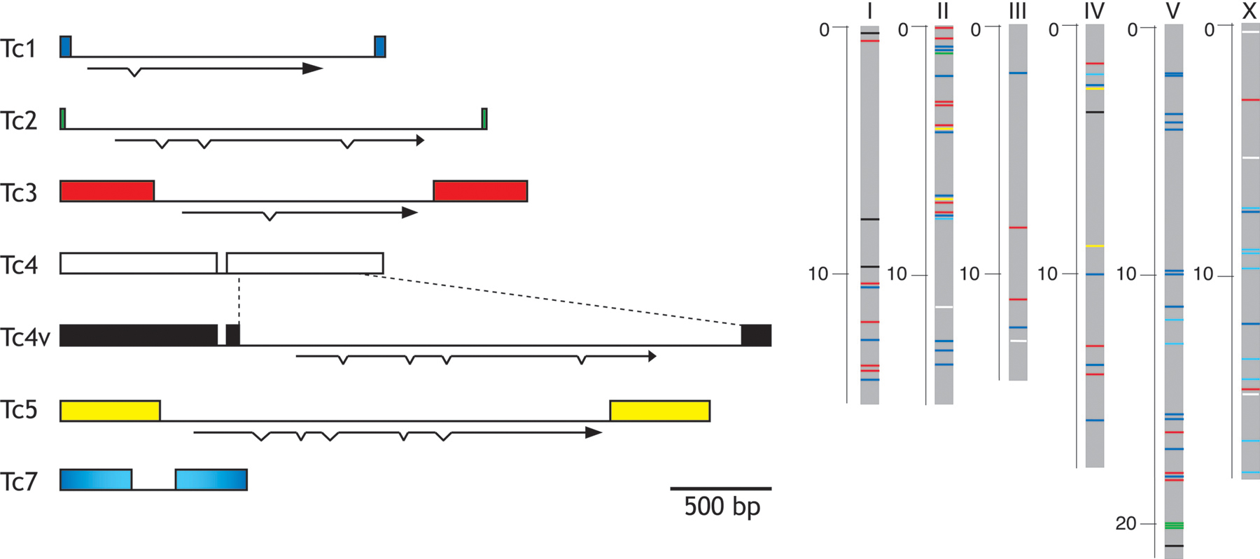
Figure 1. Active transposons in C. elegans. The structure of the transposons that proved to be mobile are depicted in the left panel. They all are class II DNA tranposons flanked by Terminal Inverted Repeats (boxes) and containing one open reading frame (arrows) encoding the transposase enzyme. Distribution of the different transposons in the genome of the C. elegans Bristol N2 strain is represented in the right panel using the same color code as on the right (Tc7 are in light blue; however, as represented on the left panel, 36 of the 38 outer bp of Tc7 are identical to the one of Tc1) The sizes of the chromosomes are in Mb. Reprinted from Fischer et al. (2003). Copyright © 2003 the Genetics Society of America.
Tc1 and Tc3 are the most active and best characterized transposons in C. elegans. The article of Anderson, Emmons and Moerman (1992) describes how circulating information between laboratories conducting simultaneous molecular and genetics approaches lead to the identification of the first transposable element in C. elegans, Tc1. Tc1 was isolated as a repeated sequence responsible for polymorphism among different strains (Emmons et al., 1983; Liao et al., 1983; Rosenzweig et al., 1983). Analysis of spontaneous and reversible mutations of the myosin heavy-chain unc-54 demonstrated the mobile nature of Tc1 (Eide and Anderson, 1985; Eide and Anderson, 1988). This feature was used to clone another muscle gene unc-22 by transposon tagging (Moerman et al., 1986; Moerman and Waterston, 1984). The subsequent characterization of additional spontaneous unc-22 mutations lead to the identification of Tc3 (Collins et al., 1989).
Tc1 is 1,610 bp long and contains two 54-bp terminal inverted repeats (TIRs; Rosenzweig et al., 1983). Tc3 is an element of 2,335 bp with 462 bp TIRs. The genome of the Bristol N2 strain contains 31 and 22 copies of Tc1 and Tc3, respectively (Fischer et al., 2003). These numbers are strain dependent. In some strain isolates such as Bergerac, Tc1 transposition is active in the germ line and each haploid genome contains up to 300-500 Tc1 copies (Egilmez et al., 1995; Emmons et al., 1983; Liao et al., 1983).
Tc1 and Tc3 are part of a superfamily of transposable elements which is named after its two best-studied members: Tc1 and the related transposon mariner which was identified in 1986 in Drosophila mauritiana (Jacobson et al., 1986). Tc1/mariner elements are probably the most widespread DNA transposons in nature and can be found in fungi, plants, ciliates and animals including ecdysozoans and vertebrates (for review see Plasterk et al., 1999). These transposons are about 1,300-2,400 bp in length, are flanked at either end by TIR and contain a single open reading frame that encodes a transposase enzyme. Tc1/mariner transposases all contain a triad of acidic residues (DDE or DDD) with a characteristic spacing which is shared by a superfamily of endonucleases (review in Haren et al., 1999, see below). Primary sequence conservation of the Tc1/mariner transposases is relatively low (about 15 % identity among the superfamily) but phylogenetic analysis suggests that all Tc1/mariner were derived from a common ancestor and might all transpose through similar mechanisms.
The Tc2 element was initially identified as a polymorphism marker (Levitt and Emmons, 1989). It is 2,074 bp in length and has perfect terminal inverted repeats of 24 bp (Ruvolo et al., 1992). There are 4 full-length Tc2 copies in the N2 genome, each flanked by a TA dinucleotide at either end. In addition, up to 300 copies of remnant Tc2 elements have been detected in the genome (Duret et al., 2000). Although not tested experimentally, gene prediction algorithms suggest that Tc2 encodes a 477 aa protein containing a DNA binding domain and a catalytic domain related to the DDE endonuclease superfamily. Transposition of Tc2 has been documented in the offspring of interstrain crosses between Bristol N2 and Bergerac BO or in a mut-2 background (Francis et al., 1995; Levitt and Emmons, 1989).
The first Tc4 element was identified as a mutagenic insertion in the gene unc-86 (Yuan et al., 1991). It is a fold-back element of 1.6 kb which contains almost perfect terminal inverted repeats of 774 bp with a 57-bp unique internal sequence. No open reading frame can be detected within Tc4. Five such Tc4 copies are present in N2 Bristol. A variant class of Tc4 (Tc4v, 5 copies in the N2 genome) contains a 2,343 bp sequence which replaces 477 bp in one of the inverted repeats (Li and Shaw, 1993). A transcript from Tc4v has been detected. It may encode a 537-aa protein which resembles transposases of the DDE superfamily. Tc4v might provide in trans the transposase required to mobilize all Tc4 elements. These elements duplicate a 3-bp target sequence TNA upon integration and are mobile in mut-2 (Yuan et al., 1991) and mut-7 (Ketting et al., 1999) mutator (mut) backgrounds.
The Tc5 element is present in four copies per haploid genome (Collins and Anderson, 1994). It is 3,171 bp long and has 491 bp long terminal inverted repeats. Tc5 and Tc4v share common features. Tc5 encodes a putative 532 amino acid transposase which is overall 33 % identical to the Tc4v transposase, Tc4 and Tc5 TIRs share a few short nucleotide sequences, and integration of Tc5 causes duplication of the same TNA trinucleotide sequence. Tc5 elements are mobile only in mut-2 (Collins and Anderson, 1994) and mut-7 (Ketting et al., 1999) backgrounds.
Tc7 is a 921 bp element that uses the Tc1A transposase for transposition (Rezsohazy et al., 1997). It is made up of two 345 bp inverted repeats separated by a unique sequence that does not contain an ORF. 36 of the 38 outer bp of Tc7 are identical to the ones of Tc1. Forced expression of Tc1A in somatic cells causes transposition of Tc7. Furthermore, Tc7 is mobile in the germ line in the same backgrounds as Tc1 such as mut-6 and mut-7 lines.
CemaT1 elements were identified in a genome search as a clade of transposons intermediate between mariner and Tc1 (Claudianos et al., 2002). They are 1,281 bp long and are flanked by two perfect 26 bp inverted repeats. 12 copies are present per haploid genome, of which 8 contain a single ORF encoding a putative 336 amino acid transposase. These elements can be excised in somatic cells and might possibly transpose in the germ line of TR403 strains (Brownlie and Whyard, 2004).
The genome of C. elegans contains several class II transposons that are not mobile under laboratory conditions. Tc3-CeIIa and CeIIb are closely related to Tc3 (Tu and Shao, 2002), Tc6 is a fold-back element (Dreyfus and Emmons, 1991; Dreyfus and Gelfand, 1999), Tc8 is related to the plant Tourist transposon (Le et al., 2001), Tc9 and Tc10 are related to Tc4(v) (Fischer et al., 2003), and the mariner-like element mle-1 is more closely related to the fly mariner elements than to the C. elegans Tc1 (Robertson and Lampe, 1995; Sedensky et al., 1994). In addition, up to 2 % of the C. elegans genome is made up from MITEs (miniature inverted-repeat transposable elements). MITEs are small non-autonomous elements that derive from transposons and are identified based on the presence of target site duplications and terminal inverted repeats (Oosumi et al., 1995; Oosumi et al., 1996; Surzycki and Belknap, 2000).
The genome of C. elegans also contains class I retrotransposons. These elements are subclassified into Long Terminal Repeat (LTR) retrotransposons that resemble retroviruses but usually lack the gene encoding the envelop protein and non-LTR retrotransposons. 124 sequences derived from Cer LTR retrotransposons, including 20 full-length elements, have been identified in the genome sequence of N2 (Bowen and McDonald, 1999; Britten, 1995; Frame et al., 2001; Ganko et al., 2001). They can be grouped in 19 families related either to the gypsy or Bel clades of retrotransposons. These LTR retrotransposons constitute 0.4 % of the C. elegans genome (Ganko et al., 2003). A thousand sequences derived from non-LTR retrotransposons can be detected in the genome and grouped into the 4 families: Rte (Youngman et al., 1996), NeSL (Malik and Eickbush, 2000), Sam and Frodo (Marin et al., 1998). These elements only constitute 0.2 % of the C. elegans genome (Zagrobelny et al., 2004) suggesting that retrotransposons have been altogether strongly counterselected in this compact genome as compared to other species such as Homo sapiens in which more than 40 % of the genome is composed of retroelement sequences. The C. elegans genome contains full length LTR and non-LTR elements but no retrotransposition events have been documented so far under laboratory conditions.
Tc1/mariner transposons move via a "cut-and-paste" mechanism: transposase binds the TIRs, catalyses excision and subsequent reinsertion into target DNA in a TA dinucleotide and leaves behind a double-strand DNA break which is repaired by the cellular machinery.
The Tc1A transposase is the only factor required in trans to mediate Tc1 transposition: forced expression of Tc1A in Bristol N2 enhances somatic transposition of Tc1 (Vos et al., 1993) and recombinant transposase purified from E. coli is capable of performing the entire transposition reaction in vitro (Vos et al., 1996). Similar evidence has been obtained for the Tc3A transposase (van Luenen et al., 1993; van Luenen et al., 1994). The Tc1A and Tc3A transposases are 343 and 329 acid long, respectively. They contain a bipartite N-terminal DNA-binding domain similar to the paired domains found in some transcription factors (van Pouderoyen et al., 1997; Watkins et al., 2004). This domain binds in a sequence-specific manner to bases located at the terminal part of the inverted repeats (position 7 to 26 in Tc1; Colloms et al., 1994; Vos and Plasterk, 1994; Vos et al., 1993). A non-specific DNA-binding domain located in more C-terminal regions of the transposase might interact with the first four bases of the TIR and the flanking TA dinucleotide, thus bringing the catalytic domain of the transposase in close proximity to the cleavage site.
Tc1A and Tc3A share a common catalytic triad of acidic residues with transposases and integrases and also with more distant enzymes that promote phosphoryltransfer reactions such as RNaseH (for review, see Haren et al., 1999). In these proteins the D,DX35E motif (two aspartates and a glutamate 35 residues away) was shown to be part of a catalytic pocket in which the acidic residues coordinate one or two magnesium or manganese ions that play a key role during transesterification reactions. Mutation of any of the DDE residues in Tc1A or Tc3A inactivate the transposase activity (van Luenen et al., 1994; Vos and Plasterk, 1994). Terminal inverted repeats are necessary and sufficient in vitro and in vivo for transposition as long as transposase is provided in trans. Within the TIRs, the first four bases of the transposon and the transposase binding sites located immediately downstream are strictly required for excision (van Luenen et al., 1994; Vos and Plasterk, 1994). However, an element only containing the 26 terminal nucleotides of Tc1 is mobilized in vitro ten times less efficiently than an element with full length TIRs (Vos et al., 1996), suggesting that the transposase binding sites contained in this terminal fragment are not sufficient for fully efficient transposition. First, additional internal sequence might be required in cis. Second, the distance between the two TIRs might be important since insertion of foreign DNA into Tc1 causes an exponential decrease of transposition frequency (Fischer et al., 1999). A similar phenomenon has been described in related transposons such as Sleeping Beauty (Izsvak et al., 2000), Himar1 (Lampe et al., 1998) and Mos1 (Lohe and Hartl, 2002) but its molecular basis is not known.
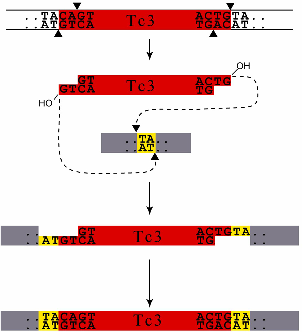
Transposon excision results from a pair of double-strand breaks at the ends of the inverted repeats (Figure 2). Coordination of cleavage at the ends of a transposon is presumably mediated by the formation of a "synaptic complex" in which the two transposon ends on the same molecule are brought together via transposase-mediated oligomerization. In Tc1 and Tc3, the 5' end is cleaved two bases from the end of the transposon and the 3' cut occurs at the junction of the transposon with the flanking DNA, resulting in a two-base pair 3'-hydroxyl overhang at each termini. Following excision, the transposon then integrates 5' of a thymidine nucleotide at a TA target sequence using the free 3' hydroxyl as a nucleophile. Repair of the resulting single-strand gaps causes a duplication of the TA dinucleotide at each transposon end. In vitro experiments demonstrated that the transposase was sufficient to catalyze excision and insertion reactions (Lampe et al., 1996; Tosi and Beverley, 2000; Vos et al., 1996). However, Tc1/mariner transposases can interact with cellular factors such as the DNA-bending protein HMGB1 which might affect the efficiency of transposition in vivo (Zayed et al., 2003).
|
Figure 2. Mechanism of Tc3 transposition. Tc3, a member of the Tc1/mariner transposon superfamily, is mobilized by a "cut-and-paste" mechanism. The transposase excises the transposon by causing double-strand breaks at the end of the transposon (arrow heads). The DNA cut is staggered, resulting in a two-base pair 3'-hydroxyl overhang at each termini. Following excision, transposons then integrates 5' of a thymidine nucleotide at a TA target sequence using the free 3' hydroxyl as a nucleophile. Repair of the resulting single-strand gaps causes a duplication of the TA dinucleotide at each transposon end. Adapted from van Luenen et al., 1994.
Tc1, Tc3 and Tc7 always integrate into the TA sequence and Tc4 and Tc5 integrate into TNA sites. Since intron sequences are AT-rich, this may explain why such elements have a higher probability of inserting into introns rather than into coding sequences (Martin et al., 2002). However, all TA dinucleotides do not represent equivalent targets. First, the difference between insertion "hot spots" and "cold spots" depends on the target sequence since in vitro transposition using naked DNA and purified Tc1A transposase mostly recapitulates Tc1 insertion preferences (Ketting et al., 1997; Vos et al., 1996). The comparison of Tc1 and Tc3 insertion sites reveals a weak consensus limited to 4 nucleotides on each side suggesting that the transposase interacts directly with the TA dinucleotide and less specifically with surrounding bases. Second, there are regional differences in insertion preferences. For example, the gene unc-22 is hit about a 100 times more frequently than unc-54 although its coding sequence is only 3.5 times larger (Eide and Anderson, 1985; Moerman and Waterston, 1984). Part of these differences might arise from the fact that transposons such as Tc1 have a preference for local reinsertion into the same chromosome from which they were excised (Fischer et al., 2003). However, Mos1, a drosophila transposon from the Tc1/mariner family was recently mobilized in C. elegans from an extrachromosomal array (Bessereau et al., 2001). Analysis of random insertions indicates the presence of a 4 kb hot-spot at the right end of chromosome I which cannot be explained by local transposition (Granger et al., 2004). Third, transposon sequences are not evenly distributed in the genome and a correlation has been observed between the density of DNA transposons but not retrotransposons and the regions of higher chromosomal recombination rate (Duret et al., 2000; Rizzon et al., 2003). Whether these two features are mechanistically linked or indirectly reflect another parameter such as differences in chromatin structure remains to be established.
Upon excision, Tc1/mariner transposons leave behind a DNA double strand break. Such breaks are repaired by the cellular machinery using two general types of repair: non homologous end joining (NHEJ) and repair by homologous recombination (for a review on DSB repair, see Haber, 2000; Figure 3). The decision to use one or another mechanism depends on tissue type and cell-cycle stage. During NHEJ, DNA ends are joined after little or no trimming. Since Tc1 and Tc3 excision leaves behind the duplicated TA and non complementary two nucleotide 5' overhangs, NHEJ generates short footprints ranging from 4 base pair insertion with TA duplications to a few base pair deletion. Occasionally, wild type sequence can be regenerated at the site of excision. NHEJ is active in C. elegans somatic cells but is also used in the germ line (Eide and Anderson, 1988; Emmons and Yesner, 1984; Emmons et al., 1983; Plasterk, 1991; Ruan and Emmons, 1987; van Luenen et al., 1994).
|
Figure 3. Repair at the excision site. Excision of a Tc1/mariner transposon generates a double-strand break in the chromosome (blue). In this example, the homologous chromosome (red) is homozygous for the Tc insertion. The double-strand break is repaired by the cellular machinery using different possible pathways. Ends can be resected, resulting in 3' single-stranded ends that can engage a recombination process. If the homologous chromosome (or the sister chromatid) is used as a template, the repair process regenerates an intact copy of the transposon at the site of excision (left panel). If resection exposes complementary strands of homologous sequences on the same chromosome (dotted ellipse), repair can occur by single-strand annealing and generates variable deletions of the sequence flanking the site of excision (middle panel). Alternatively, repair can be achieved by non-homologous end-joining (NHEJ)(right panel). In that case, ends are processed and repair leaves short footprints (dotted rectangle). For Tc1 and Tc3, one of the most common footprints contain two nucleotides from the transposon end (dark blue) and the duplicated TA.
DSB repair by homologous recombination first involves the 5' to 3' resection of the DSB ends to produce long 3'-ended single-stranded DNA tails. Several pathways can be used for repair that will generate different class of final products. In some cases, resection exposes complementary sequences allowing single-strand annealing and subsequent deletion of the segments located between these sequences. Such deletions were noticed in revertants of an unc-22 Tc1 reversion allele (Kiff et al., 1988). Zwaal et al. (Zwaal et al., 1993) demonstrated that such deletions could be isolated among the progeny of most Tc1 mutants and could be used in a reverse genetic approach to generate deletion alleles in genes of interest. Alternatively, recombination can occur with the sister chromatid or the homologous chromosome. This latter mechanism seems preferentially used for Tc1 repair in the germ line. If the animal is heterozygous for the Tc1 insertion, gene conversion will revert the broken locus to wild-type. If the animal is homozygous for the Tc1 insertion, repair will regenerate a Tc1 insertion at the excision site (Plasterk, 1991). Interestingly, analysis of Tc1 sequence polymorphisms suggest that the repair machinery can switch from allelic Tc1 template to Tc1 elements located elsewhere in the genome, resulting in chimeric Tc1 elements (Fischer et al., 2003). Occasionally, repair can be imperfect and generate partial deletions of the transposon (Fischer et al., 2003; Lohe et al., 2000).
All C. elegans strains contain numerous transposons prone to be mobilized. However, in most strains such as the reference isolate Bristol N2, transposition is only detected in somatic cells but is completely silenced in the germ line (Emmons and Yesner, 1984). In some natural isolates such as the strain Bergerac BO which was isolated in Bergerac, France (Nigon and Dougherty, 1949), Tc1 transposons are active in the germ line (Egilmez et al., 1995; Eide and Anderson, 1985; Greenwald, 1985; Moerman et al., 1986). Bergerac individuals exhibit a mutator phenotype due to spontaneous mutations caused by de novo Tc1 insertions. Strikingly, EMS-induced mutations of single loci such as mut-2 (Collins et al., 1987) or mut-7 (Ketting et al., 1999; Tabara et al., 1999) are able to globally activate the transposition of multiple Tc families including Tc1, Tc3, Tc4 and Tc5. Therefore, the germ line of C. elegans is permissive for transposition but a defense-mechanism exists to protect the genome from heritable defects caused by transposon jumps.
Transposition silencing in the germ line involves an RNA interference (RNAi)-related mechanism. This notion emerged after the realization that a set of mutants including rde-2/mut-8, rde-3/mut-2, mut-7, -14, -15 and -16 are defective for both RNAi and germ-line silencing of transposition (Chen et al., 2005; Collins et al., 1987; Ketting et al., 1999; Tabara et al., 1999; Tijsterman et al., 2002; Tops et al., 2005; Vastenhouw et al., 2003; for a complete review, see Vastenhouw and Plasterk, 2004). In a simplified model of RNAi, a double-stranded RNA (dsRNA) molecule is cleaved into 21-24 nucleotide-long short-interfering RNAs (siRNAs) by the RNAse III-like enzyme DCR-1 of the dicer family. siRNAs are loaded into the RNA-induced silencing complex (RISC) and used for specific cleavage of target RNAs. dsRNAs derived from Tc1, Tc3 and Tc5 Terminal Inverted Repeats are indeed detected in Bristol N2 animals that might arise from the fold-back of transcripts encompassing entire Tc elements. siRNAs corresponding to Tc1 and other transposons are produced in the N2 strain (Ambros et al., 2003; Sijen and Plasterk, 2003). These siRNAs seem to be functional in the germ line since a Tc1 TIR fused to gfp causes silencing of GFP expression, at least in part, by post-transcriptional silencing of the transgene in the germ line (Sijen and Plasterk, 2003). Therefore, in a simple model, RNAi might repress transposition by causing the degradation of tranposon-derived mRNA in the germ line, hence preventing the expression of any Tc transposase. In other tissues, transposon-induced RNAi might be less efficient, hence enabling somatic excision. However, mutator strains exist that are not RNAi deficient. In mut-4, -5 and -6 mutant backgrounds, transposition of Tc1 but not of other Tcs is specifically derepressed (Mori et al., 1988). These loci have not been identified at the molecular level but they have been proposed to correspond to specific Tc1 copies. For example, truncated Tc1 elements might produce transcripts which lack a sequence targeted by the RNAi surveillance system but could still produce a functional transposase. Alternatively, these elements might be full-length Tc1 elements inserted in genomic regions that lead to very efficient transcription of these copies in the germ line, hence allowing some transcripts to escape degradation. In addition to Tc1-specific mutators, a number of genes are required for global silencing of transposition but not for RNAi (Ketting et al., 1999; Vastenhouw et al., 2003). It is not clear at the moment if these genes act in a specific branch of an RNAi-dependent process or if they are involved in an RNAi-independent control of transposition.
Insertional mutagenesis with transposons generates mutant alleles that are tagged by the presence of a transposon. This molecular tag can subsequently be used to identify the mutated gene. Mutator strains and Tc1 gene tagging was used early in forward genetic screens to identify mutated-genes before the genome project reagents were available (Greenwald, 1985; Moerman et al., 1986) (for review see Anderson, 1995; Plasterk and van Luenen, 1997). Nowadays, Tc elements can be used in combination with PCR to amplify the genomic sequence that flank a mutagenic insertion and identify the mutated gene without genetic mapping (Wicks et al., 2000). However, using Tc elements as mutagens has some major drawbacks. First, the mobilization of Tc transposons is not restricted to a single class of elements in mutator strains. Second, there are several copies of each transposons in the genome which complicate the identification of the mutagenic insertion. Third, in the mutator strains that are used, transposition is not controlled. Some Tc insertions are poorly mutagenic either because they are in introns, or because they are removed from the mature mRNA by aberrant splicing (Rushforth and Anderson, 1996; Rushforth et al., 1993). Spontaneous re-excision can generate mutagenic footprints that generate a stronger phenotype but can no longer be detected in a transposon tagging strategy. These limitations have been circumvented by mobilizing the Mos1 transposon in the germ line of C. elegans (Bessereau et al., 2001). Mos1 is a member of the Tc1/mariner family and was isolated from Drosophila mauritiana (Jacobson et al., 1986). The Mos1 element is absent from the C. elegans genome and controlled mobilization of Mos1 is achieved by conditional expression of the Mos1 transposase. Mos1 mutagenesis is 10 times less efficient than chemical mutagens but the cloning of mutated genes is extremely fast since Mos1 insertions represent rare tags that are easy to localize in the genome (Gally et al., 2004; Williams et al., 2005).
Tc1 can be used to target the inactivation of a selected gene. In such approaches, Tc1 are mobilized randomly in a mutator background and independent lines are subsequently screened by PCR for the presence of a Tc1 insertion in the gene of interest (Rushforth et al., 1993). Then, deletions can be generated at low frequency by imprecise repair of double-strand breaks caused by Tc1 re-excision. The line containing the Tc1 insertion in the gene to inactivate is propagated in a mutator background. PCR analysis of the progeny can identify animals that have lost that Tc1 insertion plus a variable amount of flanking DNA (Zwaal et al., 1993). This strategy has been widely used initially, but more recent protocols of gene inactivation utilize chemical mutagens to generate random deletions in the C. elegans genome (Jansen et al., 1997). However, transposon targeted gene inactivation can still be useful when these other strategies fail or if a transposon has already been identified in the gene to inactivate (Korswagen et al., 1996; Martin et al., 2002).
Gene targeting techniques based on homologous recombination, such as those used in mice and yeast, are difficult in C. elegans (Berezikov et al., 2004; Broverman et al., 1993). Transposons might represent an interesting alternative to engineer specific mutations in the genome. In 1992, Plasterk and Groenen (Plasterk and Groenen, 1992) demonstrated that a transgene containing a fragment of the unc-22 gene could be used as a template to repair a double-strand break caused by Tc1 excision out of the unc-22 locus in a mut-6 background (Plasterk and Groenen, 1992). Point mutations contained in the transgene were copied in the genome during the repair process. Therefore, transgene-instructed gene conversion provides a strategy to engineer mutations or introduce exogenous sequence in the genome. However, such events were rare (2.10-5 event per meiosis), thus preventing this technique from being widely utilized in C. elegans. Recently, a similar strategy was tried in different mutator backgrounds to engineer deletions and introduce GFP in the genome (Barrett et al., 2004). Transgene-instructed gene conversion was found to be 10 times more frequent than previously reported, thus providing an efficient technique to engineer custom alleles in a reverse genetic approach. Efforts to generate libraries containing transposon insertions in most C. elegans genes would expand the use of this method (Granger et al., 2004; Martin et al., 2002).
I thank V. Robert and R. Weimer for critical reading of the manuscript and M. Augustin for stimulating interactions. This work was funded by an AVENIR grant from the Institut National de la Sante et de la Recherche Médicale, and by a European Union grant (6th Framework program, code NEMAGETAG).
Ambros, V., Lee, R.C., Lavanway, A., Williams, P.T., and Jewell, D. (2003). MicroRNAs and other tiny endogenous RNAs in C. elegans. Curr. Biol. 13, 807–818. Abstract Article
Anderson, P. (1995). Mutagenesis. Methods Cell. Biol. 48, 31–58. Abstract
Barrett, P.L., Fleming, J.T., and Gobel, V. (2004). Targeted gene alteration in Caenorhabditis elegans by gene conversion. Nat. Genet 36, 1231–1237. Abstract Article
Berezikov, E., Bargmann, C.I., and Plasterk, R.H. (2004). Homologous gene targeting in Caenorhabditis elegans by biolistic transformation. Nucleic Acids Res. 32, e40. Abstract Article
Bessereau, J.L., Wright, A., Williams, D.C., Schuske, K., Davis, M.W., and Jorgensen, E.M. (2001). Mobilization of a Drosophila transposon in the Caenorhabditis elegans germ line. Nature 413, 70–74. Abstract Article
Bowen, N.J., and McDonald, J.F. (1999). Genomic analysis of Caenorhabditis elegans reveals ancient families of retroviral-like elements. Genome Res. 9, 924–935. Abstract Article
Britten, R.J. (1995). Active gypsy/Ty3 retrotransposons or retroviruses in Caenorhabditis elegans. Proc. Natl. Acad. Sci. USA 92, 599–601. Abstract
Brookfield, J.F. (2005). The ecology of the genome - mobile DNA elements and their hosts. Nat. Rev. Genet. 6, 128–136. Abstract Article
Broverman, S., MacMorris, M., and Blumenthal, T. (1993). Alteration of Caenorhabditis elegans gene expression by targeted transformation. Proc. Natl. Acad. Sci. USA 90, 4359–4363. Abstract
Brownlie, J.C., and Whyard, S. (2004). CemaT1 is an active transposon within the Caenorhabditis elegans genome. Gene 338, 55–64. Abstract Article
Chen, C.-C.-G., Simard, M.J., Tabara, H., Brownell, D.R., McCollough, J.A., and Mello, C.C. (2005). A member of the polymerase beta nucleotidyltransferase superfamily is required for RNA interference in C. elegans. Curr. Biol. in press.
Claudianos, C., Brownlie, J., Russell, R., Oakeshott, J., and Whyard, S. (2002). maT--a clade of transposons intermediate between mariner and Tc1. Mol. Biol. Evol. 19, 2101–2109. Abstract
Collins, J., Forbes, E., and Anderson, P. (1989). The Tc3 family of transposable genetic elements in Caenorhabditis elegans. Genetics 121, 47–55. Abstract
Collins, J., Saari, B., and Anderson, P. (1987). Activation of a transposable element in the germ line but not the soma of Caenorhabditis elegans. Nature 328, 726–728. Abstract Article
Collins, J.J., and Anderson, P. (1994). The Tc5 family of transposable elements in Caenorhabditis elegans. Genetics 137, 771–781. Abstract
Colloms, S.D., van Luenen, H.G., and Plasterk, R.H. (1994). DNA binding activities of the Caenorhabditis elegans Tc3 transposase. Nucleic Acids Res. 22, 5548–5554. Abstract
C. elegans Sequencing Consortium. (1998). Genome sequence of the nematode C. elegans: a platform for investigating biology. Science 282, 2012–2018. Abstract Article
Dreyfus, D.H., and Emmons, S.W. (1991). A transposon-related palindromic repetitive sequence from C. elegans. Nucleic Acids Res. 19, 1871–1877. Abstract
Dreyfus, D.H., and Gelfand, E.W. (1999). Comparative analysis of invertebrate Tc6 sequences that resemble the vertebrate V(D)J recombination signal sequences (RSS). Mol. Immunol. 36, 481–488. Abstract Article
Duret, L., Marais, G., and Biemont, C. (2000). Transposons but not retrotransposons are located preferentially in regions of high recombination rate in Caenorhabditis elegans. Genetics 156, 1661–1669. Abstract
Egilmez, N.K., Ebert, R.H., II, and Shmookler Reis, R.J. (1995). Strain evolution in Caenorhabditis elegans: transposable elements as markers of interstrain evolutionary history. J. Mol. Evol. 40, 372–381. Abstract Article
Eide, D., and Anderson, P. (1985). Transposition of Tc1 in the nematode Caenorhabditis elegans. Proc. Natl. Acad. Sci. USA 82, 1756–1760. Abstract
Eide, D., and Anderson, P. (1988). Insertion and excision of Caenorhabditis elegans transposable element Tc1. Mol. Cell Biol. 8, 737–746. Abstract
Emmons, S.W., and Yesner, L. (1984). High-frequency excision of transposable element Tc 1 in the nematode Caenorhabditis elegans is limited to somatic cells. Cell 36, 599–605. Abstract Article
Emmons, S.W., Yesner, L., Ruan, K.S., and Katzenberg, D. (1983). Evidence for a transposon in Caenorhabditis elegans. Cell 32, 55–65. Abstract Article
Fischer, S.E., van Luenen, H.G., and Plasterk, R.H. (1999). Cis requirements for transposition of Tc1-like transposons in C. elegans. Mol. Gen. Genet 262, 268–274. Abstract Article
Fischer, S.E., Wienholds, E., and Plasterk, R.H. (2003). Continuous exchange of sequence information between dispersed Tc1 transposons in the Caenorhabditis elegans genome. Genetics 164, 127–134. Abstract
Frame, I.G., Cutfield, J.F., and Poulter, R.T. (2001). New BEL-like LTR-retrotransposons in Fugu rubripes, Caenorhabditis elegans, and Drosophila melanogaster. Gene 263, 219–230. Abstract Article
Francis, R., Barton, M.K., Kimble, J., and Schedl, T. (1995). gld-1, a tumor suppressor gene required for oocyte development in Caenorhabditis elegans. Genetics 139, 579–606. Abstract
Gally, C., Eimer, S., Richmond, J.E., and Bessereau, J.L. (2004). A transmembrane protein required for acetylcholine receptor clustering in Caenorhabditis elegans. Nature 431, 578–582. Abstract Article
Ganko, E.W., Bhattacharjee, V., Schliekelman, P., and McDonald, J.F. (2003). Evidence for the contribution of LTR retrotransposons to C. elegans gene evolution. Mol. Biol. Evol. 20, 1925–1931. Abstract Article
Ganko, E.W., Fielman, K.T., and McDonald, J.F. (2001). Evolutionary history of Cer elements and their impact on the C. elegans genome. Genome Res. 11, 2066–2074. Abstract Article
Granger, L., Martin, E., and Segalat, L. (2004). Mos as a tool for genome-wide insertional mutagenesis in Caenorhabditis elegans: results of a pilot study. Nucleic Acids Res. 32, e117. Abstract Article
Greenwald, I. (1985). lin-12, a nematode homeotic gene, is homologous to a set of mammalian proteins that includes epidermal growth factor. Cell 43, 583–590. Abstract Article
Haber, J.E. (2000). Partners and pathwaysrepairing a double-strand break. Trends Genet. 16, 259–264. Abstract Article
Haren, L., Ton-Hoang, B., and Chandler, M. (1999). Integrating DNA: transposases and retroviral integrases. Annu. Rev. Microbiol. 53, 245–281. Abstract Article
Izsvak, Z., Ivics, Z., and Plasterk, R.H. (2000). Sleeping Beauty, a wide host-range transposon vector for genetic transformation in vertebrates. J. Mol. Biol. 302, 93–102. Abstract Article
Jacobson, J.W., Medhora, M.M., and Hartl, D.L. (1986). Molecular structure of a somatically unstable transposable element in Drosophila. Proc. Natl. Acad. Sci. USA 83, 8684–8688. Abstract
Jansen, G., Hazendonk, E., Thijssen, K.L., and Plasterk, R.H. (1997). Reverse genetics by chemical mutagenesis in Caenorhabditis elegans. Nat. Genet. 17, 119–121. Abstract Article
Kazazian, H.H., Jr., (2004). Mobile elements: drivers of genome evolution. Science 303, 1626–1632. Abstract Article
Ketting, R.F., Fischer, S.E., and Plasterk, R.H. (1997). Target choice determinants of the Tc1 transposon of Caenorhabditis elegans. Nucleic Acids Res. 25, 4041–4047. Abstract Article
Ketting, R.F., Haverkamp, T.H., van Luenen, H.G., and Plasterk, R.H. (1999). Mut-7 of C. elegans, required for transposon silencing and RNA interference, is a homolog of Werner syndrome helicase and RNaseD. Cell 99, 133–141. Abstract Article
Kiff, J.E., Moerman, D.G., Schriefer, L.A., and Waterston, R.H. (1988). Transposon-induced deletions in unc-22 of C. elegans associated with almost normal gene activity. Nature 331, 631–633. Abstract Article
Korswagen, H.C., Durbin, R.M., Smits, M.T., and Plasterk, R.H. (1996). Transposon Tc1-derived, sequence-tagged sites in Caenorhabditis elegans as markers for gene mapping. Proc. Natl. Acad. Sci. USA 93, 14680–14685. Abstract Article
Lampe, D.J., Churchill, M.E., and Robertson, H.M. (1996). A purified mariner transposase is sufficient to mediate transposition in vitro. EMBO J. 15, 5470–5479. Abstract
Lampe, D.J., Grant, T.E., and Robertson, H.M. (1998). Factors affecting transposition of the Himar1 mariner transposon in vitro. Genetics 149, 179–187. Abstract
Le, Q.H., Turcotte, K., and Bureau, T. (2001). Tc8, a Tourist-like transposon in Caenorhabditis elegans. Genetics 158, 1081–1088. Abstract
Levitt, A., and Emmons, S.W. (1989). The Tc2 transposon in Caenorhabditis elegans. Proc. Natl. Acad. Sci. USA 86, 3232–3236. Abstract
Li, W., and Shaw, J.E. (1993). A variant Tc4 transposable element in the nematode C. elegans could encode a novel protein. Nucleic Acids Res. 21, 59–67. Abstract
Liao, L.W., Rosenzweig, B., and Hirsh, D. (1983). Analysis of a transposable element in Caenorhabditis elegans. Proc. Natl. Acad. Sci. USA 80, 3585–3589. Abstract
Lohe, A.R., and Hartl, D.L. (2002). Efficient mobilization of mariner in vivo requires multiple internal sequences. Genetics 160, 519–526. Abstract
Lohe, A.R., Timmons, C., Beerman, I., Lozovskaya, E.R., and Hartl, D.L. (2000). Self-inflicted wounds, template-directed gap repair and a recombination hotspot. Effects of the mariner transposase. Genetics 154, 647–656. Abstract
Malik, H.S., and Eickbush, T.H. (2000). NeSL-1, an ancient lineage of site-specific non-LTR retrotransposons from Caenorhabditis elegans. Genetics 154, 193–203. Abstract
Marin, I., Plata-Rengifo, P., Labrador, M., and Fontdevila, A. (1998). Evolutionary relationships among the members of an ancient class of non-LTR retrotransposons found in the nematode Caenorhabditis elegans. Mol. Biol. Evol. 15, 1390–1402. Abstract
Martin, E., Laloux, H., Couette, G., Alvarez, T., Bessou, C., Hauser, O., Sookhareea, S., Labouesse, M., and Segalat, L. (2002). Identification of 1088 new transposon insertions of Caenorhabditis elegans: a pilot study toward large-scale screens. Genetics 162, 521–524. Abstract
Moerman, D.G., Benian, G.M., and Waterston, R.H. (1986). Molecular cloning of the muscle gene unc-22 in Caenorhabditis elegans by Tc1 transposon tagging. Proc. Natl. Acad. Sci. USA 83, 2579–2583. Abstract
Moerman, D.G., and Waterston, R.H. (1984). Spontaneous unstable unc-22 IV mutations in C. elegans var. Bergerac. Genetics 108, 859–877. Abstract
Mori, I., Moerman, D.G., and Waterston, R.H. (1988). Analysis of a mutator activity necessary for germline transposition and excision of Tc1 transposable elements in Caenorhabditis elegans. Genetics 120, 397–407. Abstract
Nigon, V., and Dougherty, E.C. (1949). Reproductive patterns and attempts at reciprocal crossing of Rhabditis elegans Maupas, 1900, and Rhabditis briggsae Dougherty and Nigon, 1949 (Nematoda: Rhabditidae). J. Exp. Zool. 112, 485–503. Abstract Article
Oosumi, T., Garlick, B., and Belknap, W.R. (1995). Identification and characterization of putative transposable DNA elements in solanaceous plants and Caenorhabditis elegans. Proc. Natl. Acad. Sci. USA 92, 8886–8890. Abstract
Oosumi, T., Garlick, B., and Belknap, W.R. (1996). Identification of putative nonautonomous transposable elements associated with several transposon families in Caenorhabditis elegans. J. Mol. Evol. 43, 11–18. Abstract
Orgel, L.E., and Crick, F.H. (1980). Selfish DNA: the ultimate parasite. Nature 284, 604–607. Abstract Article
Plasterk, R.H. (1991). The origin of footprints of the Tc1 transposon of Caenorhabditis elegans. EMBO J. 10, 1919–1925. Abstract
Plasterk, R.H., and Groenen, J.T. (1992). Targeted alterations of the Caenorhabditis elegans genome by transgene instructed DNA double strand break repair following Tc1 excision. EMBO J. 11, 287–290. Abstract
Plasterk, R.H., Izsvak, Z., and Ivics, Z. (1999). Resident aliens: the Tc1/mariner superfamily of transposable elements. Trends Genet. 15, 326–332. Abstract Article
Plasterk, R.H.A., and van Luenen, H.G.A.M. (1997). Transposons. In C. elegans II,D.L. Riddle, T. Blumenthal, B.J. Meyer, and J.R. Priess, eds. (New York: Cold Spring Harbor Laboratory Press), pp. 97–116.
Rezsohazy, R., van Luenen, H.G., Durbin, R.M., and Plasterk, R.H. (1997). Tc7, a Tc1-hitch hiking transposon in Caenorhabditis elegans. Nucleic Acids Res. 25, 4048–4054. Abstract Article
Rizzon, C., Martin, E., Marais, G., Duret, L., Segalat, L., and Biemont, C. (2003). Patterns of selection against transposons inferred from the distribution of Tc1, Tc3 and Tc5 insertions in the mut-7 line of the nematode Caenorhabditis elegans. Genetics 165, 1127–1135. Abstract
Robertson, H.M., and Lampe, D.J. (1995). Recent horizontal transfer of a mariner transposable element among and between Diptera and Neuroptera. Mol. Biol. Evol. 12, 850–862. Abstract
Rosenzweig, B., Liao, L.W., and Hirsh, D. (1983). Sequence of the C. elegans transposable element Tc1. Nucleic Acids Res. 11, 4201–4209. Abstract
Ruan, K.S., and Emmons, S.W. (1987). Precise and imprecise somatic excision of the transposon Tc1 in the nematode C. elegans. Nucleic Acids Res. 15, 6875–6881. Abstract
Rushforth, A.M., and Anderson, P. (1996). Splicing removes the Caenorhabditis elegans transposon Tc1 from most mutant pre-mRNAs. Mol. Cell. Biol. 16, 422–429.
Rushforth, A.M., Saari, B., and Anderson, P. (1993). Site-selected insertion of the transposon Tc1 into a Caenorhabditis elegans myosin light chain gene. Mol. Cell Biol. 13, 902–910.
Ruvolo, V., Hill, J.E., and Levitt, A. (1992). The Tc2 transposon of Caenorhabditis elegans has the structure of a self-regulated element. DNA Cell Biol. 11, 111–122. Abstract
Sedensky, M.M., Hudson, S.J., Everson, B., and Morgan, P.G. (1994). Identification of a mariner-like repetitive sequence in C. elegans. Nucleic Acids Res. 22, 1719–1723. Abstract
Sijen, T., and Plasterk, R.H. (2003). Transposon silencing in the Caenorhabditis elegans germ line by natural RNAi. Nature 426, 310–314. Abstract Article
Stein, L.D., Bao, Z., Blasiar, D., Blumenthal, T., Brent, M.R., Chen, N., Chinwalla, A., Clarke, L., Clee, C., Coghlan, A., et al. (2003). The genome sequence ofss Caenorhabditis briggsae: a platform for comparative genomics. PLoS. Biol. 1, E45. Abstract Article
Surzycki, S.A., and Belknap, W.R. (2000). Repetitive-DNA elements are similarly distributed on Caenorhabditis elegans autosomes. Proc. Natl. Acad. Sci. USA 97, 245–249. Abstract Article
Tabara, H., Sarkissian, M., Kelly, W.G., Fleenor, J., Grishok, A., Timmons, L., Fire, A., and Mello, C.C. (1999). The rde-1 gene, RNA interference, and transposon silencing in C. elegans. Cell 99, 123–132. Abstract Article
Tijsterman, M., Ketting, R.F., Okihara, K.L., Sijen, T., and Plasterk, R.H. (2002). RNA helicase MUT-14-dependent gene silencing triggered in C. elegans by short antisense RNAs. Science 295, 694–697. Abstract Article
Tops, B.B., Tabara, H., Sijen, T., Simmer, F., Mello, C.C., Plasterk, R.H., and Ketting, R.F. (2005). RDE-2 interacts with MUT-7 to mediate RNA interference in Caenorhabditis elegans. Nucleic Acids Res. 33, 347–355. Abstract Article
Tosi, L.R., and Beverley, S.M. (2000). cis and trans factors affecting Mos1 mariner evolution and transposition in vitro, and its potential for functional genomics. Nucleic Acids Res. 28, 784–790. Abstract Article
Tu, Z., and Shao, H. (2002). Intra- and inter-specific diversity of Tc3-like transposons in nematodes and insects and implications for their evolution and transposition. Gene 282, 133–142. Abstract Article
van Luenen, H.G., Colloms, S.D., and Plasterk, R.H. (1993). Mobilization of quiet, endogenous Tc3 transposons of Caenorhabditis elegans by forced expression of Tc3 transposase. EMBO J. 12, 2513–2520. Abstract
van Luenen, H.G., Colloms, S.D., and Plasterk, R.H. (1994). The mechanism of transposition of Tc3 in C. elegans. Cell 79, 293–301. Abstract Article
van Pouderoyen, G., Ketting, R.F., Perrakis, A., Plasterk, R.H., and Sixma, T.K. (1997). Crystal structure of the specific DNA-binding domain of Tc3 transposase of C. elegans in complex with transposon DNA. EMBO J. 16, 6044–6054. Abstract Article
Vastenhouw, N.L., Fischer, S.E., Robert, V.J., Thijssen, K.L., Fraser, A.G., Kamath, R.S., Ahringer, J., and Plasterk, R.H. (2003). A genome-wide screen identifies 27 genes involved in transposon silencing in C. elegans. Curr. Biol. 13, 1311–1316. Abstract Article
Vastenhouw, N.L., and Plasterk, R.H. (2004). RNAi protects the Caenorhabditis elegans germline against transposition. Trends Genet 20, 314–319. Abstract Article
Vos, J.C., De Baere, I., and Plasterk, R.H. (1996). Transposase is the only nematode protein required for in vitro transposition of Tc1. Genes Dev. 10, 755–761. Abstract
Vos, J.C., and Plasterk, R.H. (1994). Tc1 transposase of Caenorhabditis elegans is an endonuclease with a bipartite DNA binding domain. EMBO J. 13, 6125–6132. Abstract
Vos, J.C., van Luenen, H.G., and Plasterk, R.H. (1993). Characterization of the Caenorhabditis elegans Tc1 transposase in vivo and in vitro. Genes Dev. 7, 1244–1253. Abstract
Watkins, S., van Pouderoyen, G., and Sixma, T.K. (2004). Structural analysis of the bipartite DNA-binding domain of Tc3 transposase bound to transposon DNA. Nucleic Acids Res. 32, 4306–4312. Abstract Article
Wicks, S.R., de Vries, C.J., van Luenen, H.G., and Plasterk, R.H. (2000). CHE-3, a cytosolic dynein heavy chain, is required for sensory cilia structure and function in Caenorhabditis elegans. Dev. Biol. 221, 295–307. Abstract Article
Williams, D.C., Boulin, T., Ruaud, A.F., Jorgensen, E.M., and Bessereau, J.L. (2005). Characterization of Mos1 Mediated Mutagenesis in C. elegans: A Method for the Rapid Identification of Mutated Genes. Genetics. Abstract Article
Youngman, S., van Luenen, H.G., and Plasterk, R.H. (1996). Rte-1, a retrotransposon-like element in Caenorhabditis elegans. FEBS Lett. 380, 1–7. Abstract Article
Yuan, J.Y., Finney, M., Tsung, N., and Horvitz, H.R. (1991). Tc4, a Caenorhabditis elegans transposable element with an unusual fold-back structure. Proc. Natl. Acad. Sci. USA 88, 3334–3338. Abstract
Zagrobelny, M., Jeffares, D.C., and Arctander, P. (2004). Differences in non-LTR retrotransposons within C. elegans and C. briggsae genomes. Gene 330, 61–66. Abstract Article
Zayed, H., Izsvak, Z., Khare, D., Heinemann, U., and Ivics, Z. (2003). The DNA-bending protein HMGB1 is a cellular cofactor of Sleeping Beauty transposition. Nucleic Acids Res. 31, 2313–2322. Abstract Article
Zwaal, R.R., Broeks, A., van Meurs, J., Groenen, J.T., and Plasterk, R.H. (1993). Target-selected gene inactivation in Caenorhabditis elegans by using a frozen transposon insertion mutant bank. Proc. Natl. Acad. Sci. USA 90, 7431–7435. Abstract
*Edited by Jonathan Hodgkin and Philip Anderson. Last revised April 5, 2005. Published January 18, 2006. This chapter should be cited as: Bessereau, J.-L. Transposons in C. elegans (January 18, 2006), WormBook, ed. The C. elegans Research Community, WormBook, doi/10.1895/wormbook.1.70.1, http://www.wormbook.org.
Copyright: © 2006 Jean-Louis Bessereau. This is an open-access article distributed under the terms of the Creative Commons Attribution License, which permits unrestricted use, distribution, and reproduction in any medium, provided the original author and source are credited.
§To whom correspondence should be addressed. E-mail: jlbesse@biologie.ens.fr
 All WormBook content, except where otherwise noted, is licensed under a Creative Commons Attribution License.