Embryo series courtesy of Einhard Schierenberg
Table of Contents
Abstract
Caenorhabditis elegans has orthologs for most of the key enzymes involved in eukaryotic intermediary metabolism, suggesting that the major metabolic pathways are probably present in this species. We discuss how metabolic patterns and activity change as the worm traverses development and ages, or responds to unfavorable external factors, such as temperature extremes or shortages in food or oxygen. Dauer diapause is marked by an enhanced resistance to oxidative stress and a shift toward microaerobic and anaplerotic metabolic pathways and hypometabolism, as indicated by the increased importance of the malate dismutation and glyoxylate pathways and the repression of citric acid cycle activity. These alterations promote prolonged survival of the dauer larva; some of these changes also accompany the extended lifespan of insulin/IGF-1 and several mitochondrial mutants. We also present a brief overview of the nutritional requirements, energy storage and waste products generated by C. elegans.
Metabolism comprises the entire set of chemical reactions that occur in a living organism that allow it to reproduce, develop, maintain its structure and respond to the environment. These chemical reactions form an intricate network of pathways and cycles in which the flow of reaction products (metabolites) is determined by many regulatory mechanisms. Traditionally, metabolism is subdivided into catabolism, the breaking down of complex molecules, and anabolism, processes related to the synthesis of complex organic substances.
According to the definition provided above, metabolism includes every cellular process, ranging from DNA replication to transcription and translation to enzyme function, and also involves the chemistry of small molecules in the cell. In this chapter, we will focus on intermediary metabolism, which describes all reactions concerned with the storage and generation of metabolic energy required for the biosynthesis of low-molecular weight compounds and energy storage compounds (Mathews and Van Holde, 1996). In the intermediary metabolism pathway, the structure of each enzyme plays a crucial role in determining the specific properties of each reaction.
In the early seventies, Brenner introduced Caenorhabditis elegans as a multicellular genetic model, mainly because this organism has very few somatic cells, which made it possible for investigators to reconstruct the cell lineage and to map the wiring of the nervous system (Brenner, 1974). Since then, it has become one of the most powerful tools for studying the genetics of metazoan development, neurobiology, aging and many other biological processes. This worm, however, is less amenable to classical biochemical study. It is sometimes difficult to obtain large quantities of synchronized individuals; eggshells and cuticles are tough barriers and it is virtually impossible to collect pure tissue in biochemically relevant quantities (Mains and McGhee, 1999). Nevertheless, many metabolic studies of free-living and parasitic nematodes (Bolla, 1980) have allowed us to gain deeper insights into the complex biochemical events that underpin the life of C. elegans.
The intermediary metabolic network is well conserved among eukaryotes and the major metabolic pathways found in heterotrophic organisms are also present in C. elegans (Vastrik et al., 2007). In the first section of this chapter, we will elaborate on the characteristics of these general pathways in our C. elegans model. We will subsequently focus on the vital compounds that cannot be synthesized by C. elegans and hence need to be extracted from the environment. Vitamins, essential amino acids and other related compounds will be discussed in the section about nutritional requirements. At the end of this chapter, a short overview will also be given describing the different metabolic waste products and the storage and production of metabolic energy in C. elegans.
At many intersections within the metabolic network, the flow of metabolites can be diverted to a specific direction according to the need of the organism at that particular moment. These metabolic shifts can occur instantly, e.g. as a response to changing environments, or more slowly over the course of the C. elegans life cycle. In Section 6, we will explain some of the typical metabolic alterations that take place from embryogenesis to the last larval stage, including the specialized dauer stage. We will also focus on the metabolic changes that accompany the worm aging process, and the large fluctuations in biotic and abiotic environmental factors that might be encountered by the soil dwelling C. elegans. Finally, we will discuss the metabolic patterns that arise due to changes in temperature, food restriction, oxygen deprivation or osmotic stress.
Recent estimates for the last common ancestor to vertebrates and nematodes range from 800 million to over one billion years ago (Hedges and Kumar, 2003; Hedges, 2002; Ureta-Vidal et al., 2003; Blaxter, 1998). Since that time, nematodes have developed a bewildering variety of life histories including parasitism of animals and plants and the ability to live in harsh environments. Conceivably, conserved eukaryotic metabolic pathways are more likely to be found in free-living bacterivorous nematodes such as C. elegans, which live under mostly temperate conditions. During the 1960s and -70s there was much interest in comparative biochemistry and a great deal of work was done on the metabolism of nematodes, albeit predominantly focusing on parasitic forms (Bolla, 1980; Atkinson, 1980). The other free-living species studied most intensively included the Pangrolaimidae Panagrellus redivivus and Turbatrix aceti, and a Rhabditidae, Caenorhabditis briggsae. C. elegans and C. briggsae are sibling species, which display only a few subtle morphological differences. Their genomes have been completely sequenced and approximately 62% of the predicted C. briggsae genes are one-to-one orthologs of C. elegans (Stein et al., 2003). Thus there is little doubt that biochemical reactions identified in C. briggsae also occur in C. elegans and vice versa.
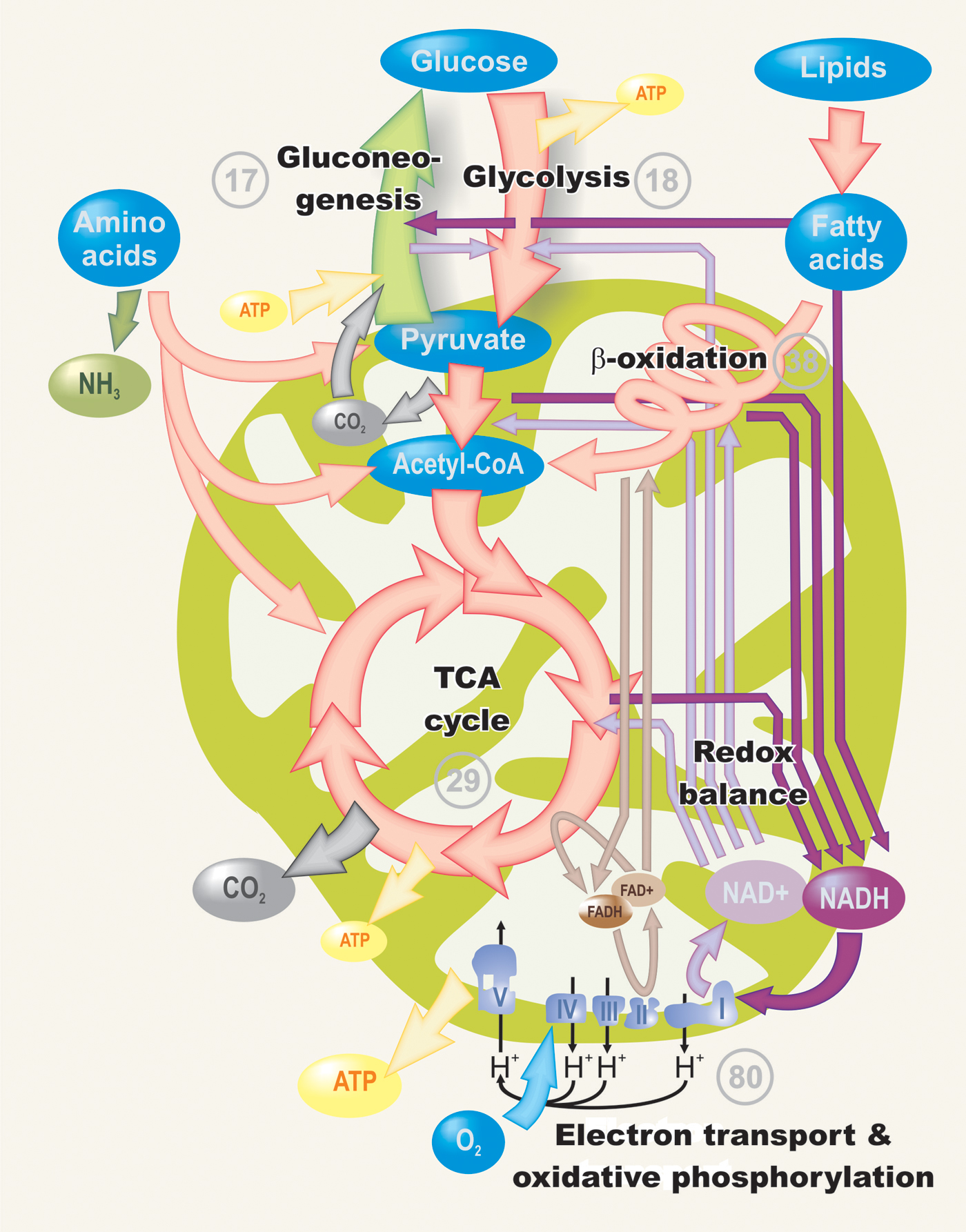
Databases such as Wormbase, Reactome and KEGG show that C. elegans orthologs have been found for most of the enzymes involved in the main pathways of intermediary metabolism. Of course, this only provides information about the metabolic machinery present in the animal; the activities of these biochemical pathways can only be determined by gene expression and enzyme studies and by quantifying metabolites. The number of C. elegans orthologs that are currently identified is indicated on the diagram representing the major metabolic pathways, which will be discussed later in this chapter (Figure 1).
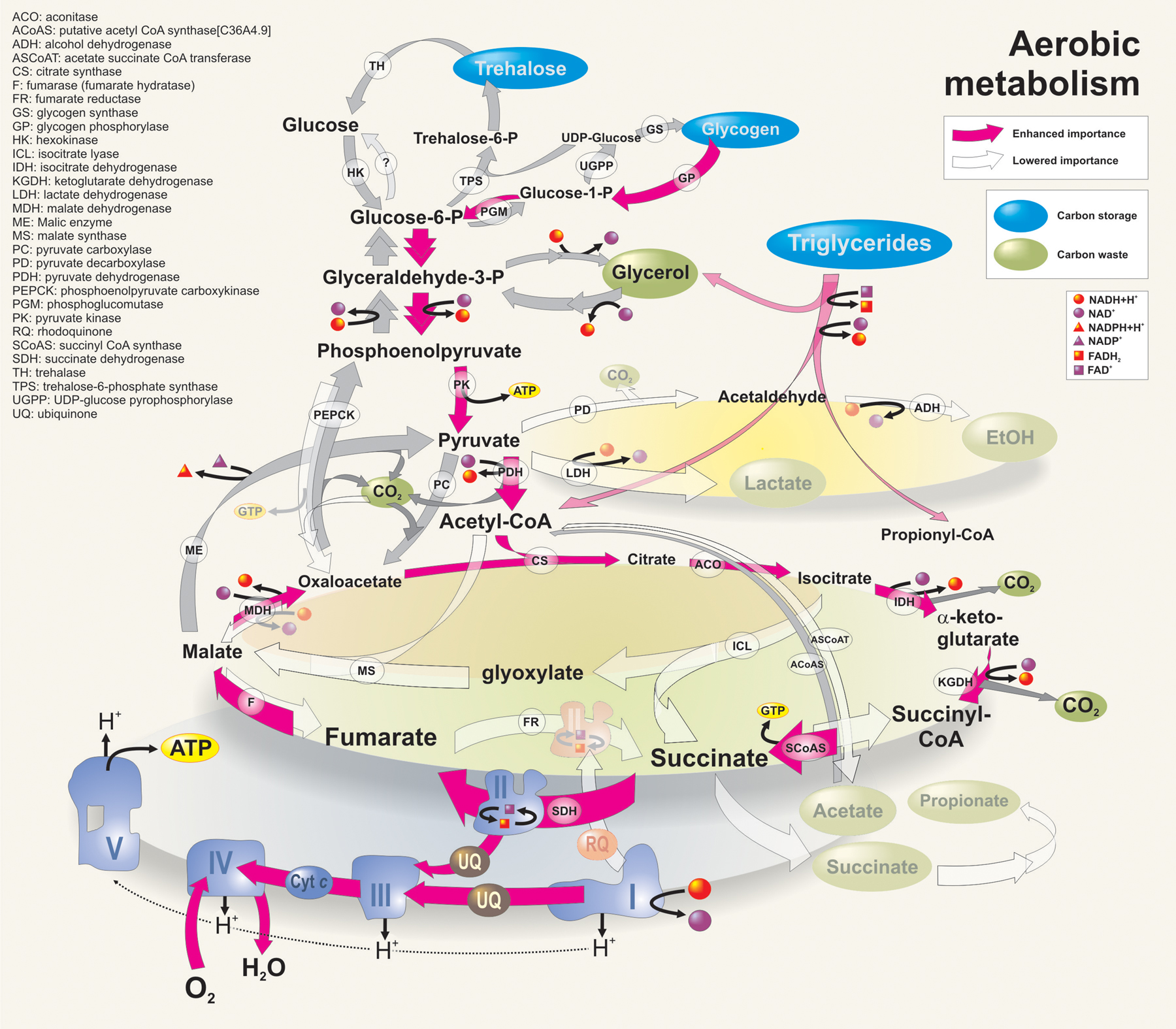
Under aerobic conditions (Figure 2), C. elegans will metabolize energy through the standard metabolic pathways. In glycolysis, a series of enzymatic reactions will convert sugars (typically glucose) to pyruvate. During this process, small amounts of ATP are generated and electron carriers are loaded with electrons (NAD+ is reduced to NADH + H+). Consequently, pyruvate is translocated to the mitochondria, decarboxylated and converted into acetyl-CoA. In this process the first completely oxidized carbon is released as CO2. Acetyl-CoA then enters the tricarboxylic acid (TCA) cycle by condensing with oxaloacetate to form citrate. A series of oxidation reactions then ensue with the result that two carbons are expelled as CO2, (G/A)TP is produced and the electron carriers (NAD+ and FAD+) are reduced. Finally, the cycle is completed when oxaloacetate is formed. The electron carriers that were reduced during glycolysis and the TCA cycle deliver their electrons to O2 (with the formation of water) through a series of electron transport chain redox proteins located in the inner mitochondrial membrane. During electron transfer, some of these redox proteins shuttle protons from the mitochondrial matrix to the intramembrane space of the mitochondria, thereby creating an electrochemical gradient. The potential energy in this gradient is finally used by ATP-synthase (another protein complex in the inner mitochondrial membrane) to drive ATP synthesis.
Lipids and amino acids can also be used as energy sources, but they enter the main pathways at different points. C. elegans has a functional methylmalonyl-CoA epimerase (racemase) that is involved in propionyl-CoA metabolism for the degradation of branched amino acids and odd-chain fatty acids (Kühnl et al., 2005). Fatty acid moieties of lipids are broken down by β-oxidation into acetyl-CoA (which in turn can enter the TCA cycle). β-oxidation occurs in the mitochondrial matrix and also yields reduced electron carriers. Peroxisomal β-oxidation of long-chain fatty acids is not linked directly to energy metabolism because the reduced electron carrier is directly oxidized by molecular oxygen (yielding hydrogen peroxide). Amino acids can be broken down via distinct pathways and their carbon skeletons can be metabolized in the TCA cycle.
Besides these well-known pathways of intermediary metabolism, C. elegans can use alternative pathways under distinct developmental or environmental conditions, as will be discussed in more detail in the following sections.
|
Figure 1. General overview of the major conserved pathways of intermediary metabolism in C. elegans. The numbers next to each pathway represent the number of orthologs that were detected in the C. elegans genome (based on McElwee et al., 2006).
The search for a chemically defined medium that would support sustained population growth of C. briggsae led to the formulation of a mixture, designated EM1, which was based on the amino acid ratios found in E. coli (Sayre et al., 1963). This medium was later modified by Buecher et al. (1966) and called Caenorhabditis briggsae Maintenance Medium (CbMM). This medium contained a total of 53 components consisting of minerals, glucose, amino acids, vitamins, growth factors and precursors for nucleic acid synthesis (adjusted to pH 5.9 with KOH). Caenorhabditis elegans Maintenance Medium (CeMM) has the same basal composition, but contains either more glucose or potassium acetate for energy (Lu and Goetsch, 1993; Szewczyk et al., 2003). This basal medium, which can be replaced by 3% soy peptone and 3% yeast extract, must be further supplemented with sterol and a heme source.
|
Figure 2. Metabolic flux in aerobic conditions. In the diagram, only the formation of (G)ATP is indicated (not its consumption). This diagram is primarily based on the work of McElwee et al. (2006), Holt and Riddle (2003) and Burnell et al., (2005).
C. elegans and all other nematodes studied to date lack the capacity to synthesize sterols de novo from acetate (Rothstein, 1968; Cole and Dutky, 1969). This requirement can be met by providing exogenous cholesterol, β-sitosterol, 7-dehydrocholesterol, ergosterol, or stigmasterol (Hieb and Rothstein, 1968; Szewczyk et al., 2003). Indeed, C. elegans can use various dietary sterols, including plant sterols (Chitwood and Lusby, 1991). P. redivivus and Caenorhabditis species have the capacity to synthesize lanosterol from squalene 2,3-oxide; the major metabolic block in the pathway of de novo sterol biosynthesis likely occurs between farnesol (and any other steps before farnesol) and squalene (Willett and Downey, 1974; Lu et al., 1977). More recently, Lee et al. (2005) constructed a C. elegans strain expressing human dehydrocholesterol reductase, which enabled the worms to convert exogenous 7-dehydrocholesterol into cholesterol. This transgenic strain was slightly longer-lived and stress-resistant, but showed reduced fecundity.
Sterol-supplemented chemically defined medium will not sustain nematode population growth unless a heme source is added. Hemin chloride can be used, but various heme proteins have proven more effective (Hieb et al., 1970; Vanfleteren, 1974). Hemoglobin or myoglobin is most often added (Föll et al., 1999; Murfitt et al., 1976; Houthoofd et al., 2002a; Houthoofd et al., 2003); cytochrome c can be used to prepare a medium that is free of precipitate (Szewczyk et al., 2003). C. elegans lacks orthologs of all 7 enzymes (δ-aminolevulinic acid dehydratase, porphobilinogen deaminase, uroporphyrinogen III synthase, uroporphyrinogen decarboxylase, coproporphyrinogen oxidase, protoporphyrinogen oxidase and ferrochelatase), which are needed to synthesize heme from δ-aminolevulinic acid (Rao et al., 2005).
A requirement for several vitamins has been shown: thiamin, riboflavin, folic acid, niacinamide, pantothenic acid, pyridoxine (Dougherty et al., 1959; Nicholas et al., 1962; Dougherty and Hansen, 1957; Lu et al., 1974), biotin (Nicholas and Jantunen, 1963) and vitamin B12 (Lu et al., 1976). The following essential amino acids are required to sustain population growth of C. briggsae and C. elegans: arginine, histidine, lysine, tryptophan, phenylalanine, methionine, threonine, leucine, isoleucine and valine (Vanfleteren, 1980), although the animals can synthesize limited quantities from labeled precursors (Nicholas et al., 1960; Rothstein and Tomlinson, 1961; 1962; Rothstein, 1963; 1965). By contrast, alanine, aspartic acid, cysteine, glutamic acid, glutamine, glycine, serine, and tyrosine are not essential nutrients (Vanfleteren, 1980).
In addition to long-chain fatty acids, ethanol, n-propanol and acetate also promote population growth of C. briggsae in chemically defined medium (Lu et al., 1978). These compounds are utilized as energy sources by Caenorhabditis, since comparable results were obtained when they were replaced with various carbohydrate supplements. Lu and Goetsch (1993) found that glucose and glycogen, followed by trehalose, fructose and sucrose can be utilized as energy sources. Cooper and Van Gundy (1971) reported that C. elegans is capable of aerobic, but not anaerobic, catabolism of ethanol. Di- and tricarboxylic acids can probably be transported from the extracellular environment into the gut cells and used as a fuel source in the TCA cycle (Fei et al., 2004).
Ammonia is the main excretory product of nitrogen metabolism, but nitrogen is also excreted in the form of amino acids (Rothstein, 1963; 1965). Rothstein (1965) identified several amino acids as excretion products of axenic populations of C. briggsae after they had been cultured on various radioactive precursors; the concentration of excreted amino acids was greater than the concentration retained in the tissues. Wright (1975a; 1975b) found evidence for a functional urea cycle of low activity, and excretion of urea in P. redivivus.
In vertebrates, carbon atoms of carbohydrates, lipids and amino acids are usually completely oxidized to CO2 under aerobic conditions. Carbon dioxide is also a major waste product in C. elegans but, like many other invertebrates and micro-organisms, the worm can also excrete other types of carbon waste. Rothstein (1969) and Liu and Rothstein (1976) found that C. briggsae excretes substantial amounts of glycerol as a major radioactive product when incubated with 2-14C-acetate in complete nutritional medium, but in unsupplemented buffer solution little or no glycerol was produced. The advantage of glycerol excretion in a nutrient-rich environment is not clear. Possibly it is a wasteful means to dispose of superfluous products of metabolism in a nutrient-rich environment, much like synthesis and excretion of amino acids to dispose of end products of nitrogen metabolism. This is consistent with the observation that C. elegans and C. briggsae are typical opportunistic species that need dense bacterial populations to maintain rapid growth and fecundity. Rapid excretion of glycerol to the external medium is also associated with recovery from hypertonic stress (see Section 7.4). Like other soil nematodes, C. elegans is hyperosmotic with its normal environment. The animals accumulate and excrete glycerol as needed to maintain cellular osmotic homeostasis (Lamitina et al., 2004; Wheeler and Thomas, 2006).
Free-living nematodes have the ability to synthesize saturated and unsaturated fatty acids de novo (Rothstein, 1970), and they typically store energy as lipids. Recently, it was found that most lipid species in C. elegans (triglycerides, phospholipids, sphingolipids, etc.) are composed of fatty acids directly absorbed from the bacterial food source (C. Perez and M. Van Gilst, personal communication). Up to 35% of the dry body mass of C. elegans is lipid, and triacyl glyceride fat stores (~40-55% of total lipids) are the major energy storage molecules (Cooper and Van Gundy, 1971; Lee and Atkinson, 1976; see Obesity and the regulation of fat metabolism). Carbohydrate stores consist primarily of glycogen (3.3% of the dry body mass, Cooper and Van Gundy, 1970), but significant amounts of trehalose and glucose are also present (Föll et al., 1999; Hanover et al., 2005). Interestingly, O-GlcNac transferase (OGT) modulates the relative use of these macronutrient storage molecules. OGT knockout mutants have a 3-fold elevation of trehalose levels and glycogen stores with a concomitant 3-fold decrease in triglyceride levels (Hanover et al., 2005). Cooper and van Gundy (1970) observed that the Caenorhabditis species rapidly metabolized glycogen when starved. Dauer larvae rely on their lipid reserves for long-term survival (O'Riordan and Burnell, 1990).
Biochemical studies have provided evidence for a complete tricarboxylic acid cycle in T. aceti (Rothstein et al., 1970) and C. elegans (Murfitt et al., 1976). When C. elegans encounters adverse conditions, the flow of glycolytic products to the TCA cycle may be prevented by pyruvate dehydrogenase kinase (a negative regulator of PDH) activity. Thus, this enzyme may act as a molecular switch for energy metabolism, like it does in hibernating mammals (E-J. Shin, J-E. Hwang and Y-K. Paik, personal communication). Evidence for the occurrence of the phosphogluconate pathway (also called hexose monophosphate shunt or pentose phosphate pathway) has been reported for T. aceti. This pathway oxidizes glucose to generate pentoses, CO2 and reduced NADP and is important for maintaining the cellular redox balance. Interestingly, this pathway produces free ribitol that is excreted (Panagides and Rothstein, 1973). The advantage to the animals of ribitol production and excretion is not clear. One possible explanation is that it serves as a regulatory mechanism of NADPH production although ribitol was not excreted by C. briggsae.
Nematodes, including C. elegans, also possess the glyoxylate cycle enzymes, which convert two acetyl-CoA units into succinate and malate (Rothstein and Mayoh, 1964a; 1964b; Rothstein and Mayoh, 1965; 1966; Colonna and McFadden, 1975; Patel and McFadden, 1977). Key enzymes are isocitrate lyase, which cleaves isocitrate to succinate and glyoxylate, and malate synthase, which condenses glyoxylate with acetyl-CoA to form malate. C. elegans has only one bifunctional glyoxylate cycle protein, which contains separate domains for both enzyme activities on a single polypeptide (Liu et al., 1995). In plants, the glyoxylate cycle operates in specialized peroxisomes, called glyoxysomes. The malate produced in glyoxysomes can be converted to oxaloacetate, completing the cycle. Alternatively, succinate is either transported to the mitochondria where it is converted to malate and thereby consumed in the citric acid cycle, making the glyoxylate cycle an anaplerotic (refilling) process, or transported to the cytosol and further oxidized to oxaloacetate for entry into gluconeogenesis (net production of carbohydrate from triacylglycerols) (Tolbert, 1981).
Analysis of subcellular fractions from C. elegans (Patel and McFadden, 1977) and T. aceti (Aueron and Rothstein, 1974) has also detected the presence of glyoxysome-like bodies in these species. On the other hand, glyoxylate enzymes have been reported in mitochondrial fractions from T. aceti (McKinley and Trelease, 1978a; 1978b) and Ascaris suum larvae (Rubin and Trelease, 1976).
Metabolic rate measured as oxygen consumed or heat produced per unit time and normalized to protein content exhibits life-cycle dependent modulation: it is higher in L1 larvae than in developing eggs and it further increases to reach the highest values in L2-L4 larvae, followed by a steep decrease as worms reach adulthood. The metabolic rate then gradually decreases as the worms traverse this final stage of the life cycle toward senescence (De Cuyper and Vanfleteren, 1982; Vanfleteren and De Vreese, 1996; Houthoofd et al., 2002b). The low oxygen consumption rates of developing eggs and freshly hatched larvae are likely related to the shift in energy metabolism away from the TCA cycle. In embryos, the high activity of the glyoxylate cycle is necessary for converting stored lipids so that they can be used for energy production and gluconeogenesis. After hatching, the activity of isocitrate lyase progressively decreases, whereas energy production through the TCA cycle increases substantially (Wadsworth and Riddle, 1989). Interestingly Liu et al. (1997) reported that glyoxylate cycle mRNA and protein increased during early development, peaked at the L2 stage and declined afterwards. Starvation induced glyoxylate cycle expression throughout juvenile development (Khan and McFadden, 1982; Liu et al., 1997). However, in L2 predauers and in dauer larvae, which do not feed, the glyoxylate cycle enzyme activities do not increase but closely parallel those from L2 (Wadsworth and Riddle, 1989), suggesting that the regulation of glyoxylate cycle protein expression in dauers is different from that during normal development (Liu et al., 1997). The ATP profiles reflect these changes in energy production (Houthoofd et al., 2002b). NMR spectra showed that ATP was below detection limits in L1 larvae and dauers, whereas AMP was detectable throughout development and was particularly high in L1 larvae (Wadsworth and Riddle, 1989).
Movement accounts for about 37% of the total energy expenditure of L4 larvae and this fraction decreases further to 25% in adult animals (Vanfleteren and De Vreese, 1996).
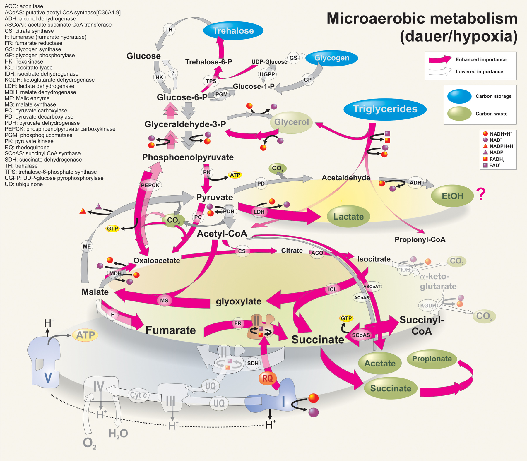
The dauer stage is an alternative third stage larva that is formed under unfavorable conditions, and is specialized for survival and dispersal (see Dauer). Since dauers do not feed, they must metabolize stored macronutrients, mainly lipids, but also glycogen and trehalose, and suppress energy-consuming metabolic activities. This is indeed the case; dauers consume less oxygen when compared to other juvenile stages (O'Riordan and Burnell, 1989; Vanfleteren and De Vreese, 1996; Houthoofd et al., 2002b) and produce less heat (Houthoofd et al., 2002b), consistent with an overall reduction of metabolic rate. This appears to be caused by the reduced activities of the enzymes involved in the glycolytic, gluconeogenic, TCA cycle and oxidative phosphorylation pathways in dauers, relative to those of adults (O'Riordan and Burnell, 1989). Micro-array and SAGE analyses of dauer larvae versus recovered dauers or mixed stage populations suggested that the transcription of enzymes involved in the β-oxidation of fatty acids, glycolysis, glyoxylate and gluconeogenesis pathways was enhanced, indicating that triglycerides were being converted into sugars (Wang and Kim, 2003; McElwee et al., 2004; 2006; Jones et al., 2001; Holt and Riddle, 2003). The flux of metabolites through the glyoxylate cyle is relatively more important in the dauer, although this shift is mainly due to a substantial decrease of the carbon flux through the TCA cycle rather than an increased flux through the glyoxylate cycle (O'Riordan and Burnell, 1990). Phosphoenolpyruvate-carboxykinase (PEPCK) levels are relatively high in the dauer, indicating that gluconeogenesis is elevated, but glycogen re-synthesis is apparently suppressed, as indicated by the relatively low activity of fructose 1,6-bisphosphatase (O'Riordan and Burnell, 1989). Thus, the high level of PEPCK activity in dauers is more likely to be associated with the fixation of CO2 into phosphoenolpyruvate to form oxaloacetate, which can be converted to malate and used in other pathways, thereby providing a useful anaplerotic mechanism for the starving dauer larva. The conversion of lipids into sugars may also be required for transportation of energy from lipid-containing cells (intestine and epidermis) into other tissues (muscle, neurons) (Ogg et al., 1997). Since the C. elegans genome does not contain a functional glucose-6-phosphatase, it is assumed that trehalose, rather than glucose is used as a transport sugar (McElwee et al., 2004) (Figure 3).
It is not clear whether ethanol fermentation is upregulated in the dauer. SAGE analyses detected enhanced transcription of the gene K12G11.3 that encodes a protein with high similarity to sorbitol dehydrogenase (which catalyzes the reversible oxidation-reduction reaction between fructose and sorbitol) and alcohol dehydrogenases from other species (Holt and Riddle, 2003). However, it has not been shown that the K12G11.3 protein is involved in ethanol fermentation. Microarray analysis detected the upregulation of two alcohol dehydrogenase genes and the downregulation of a third one (McElwee et al., 2006). Lactate dehydrogenase expression was lower in dauers, either compared to larvae 12 hours after the onset of dauer recovery (McElwee et al., 2006) or mixed-stage worms (Holt and Riddle, 2003). Thus there is little evidence that pyruvate fermentation is upregulated in the dauer.
Another anaerobic process that seems to be important in dauers is malate dismutation. Malate, formed from oxaloacetate by malate dehydrogenase or produced by the glyoxylate pathway, can enter mitochondria for aerobic (TCA cycle) or anaerobic combustion (Figure 3). Here, malate can be oxidized to pyruvate via malic enzyme and further converted into acetyl-CoA and acetate. Alternatively, malate can be reduced to fumarate and succinate, using the reverse action of fumarase and complex II of the electron transport chain. Complex II can act as succinate dehydrogenase or as fumarate reductase (Figure 3). In addition to the electron transporter ubiquinone, dauers also contain rhodoquinone, which has a lower reduction potential and favors the reverse action of complex II (Takamiya et al., 1999; Holt and Riddle, 2003; Burnell et al., 2005). Oxidation of malate to acetate and reduction to succinate in a ratio of 1:2 would maintain the redox balance in equilibrium in the absence of oxygen (Burnell et al., 2005). The malate dismutation pathway and the excretion of succinate, propionate and acetate has been well documented for parasitic helminths, including A. suum (Thielens et al., 2002). These end products of anaerobic mitochondria are also excreted by C. elegans under anoxic conditions (Föll et al., 1999).
|
Figure 3. Metabolic flux in microaerobic conditions. In the diagram, only the formation of (G)ATP is indicated (not its consumption). This diagram is primarily based on the work of McElwee et al. (2006), Holt and Riddle (2003), and Burnell et al., (2005). Note that LDH activity is increased in hypoxia only; LDH expression level is lowered in the dauer stage.
Dauers are very long-lived and it has been shown that the adult lifespan of C. elegans is independent of the time spent as a dauer larva (Klass and Hirsh, 1976). Dauers, however, show time-dependent metabolic changes, including a reduced superoxide production rate potential (as defined in Braeckman et al., 2002a), accumulation of lipofuscin and oxidized flavins and a gradual decline in aconitase activity (Houthoofd et al., 2002b). Thus, dauers seem to age slowly, but the changes accompanying the process can be reversed when the dauers develop to the L4 and adult stages. The long lifespan of dauers (up to 8 times the 2-week adult lifespan) is associated with increased protection against molecular damage (Honda and Honda, 1999; Taub et al., 1999; Birnby et al., 2000; Jones et al., 2001; Yu and Larsen, 2001; Houthoofd et al., 2002b; McElwee et al., 2003; Wang and Kim, 2003; McElwee et al., 2004; Gems and McElwee, 2005).
Notwithstanding its short lifespan, C. elegans shows clear morphological and behavioral signs of aging (Epstein et al., 1972; Duhon and Johnson, 1995; Bolanowski et al., 1981; Herndon et al., 2002). Surprisingly, the relative transcriptional and translational profiles do not change dramatically during the aging process in wild-type C. elegans (Lund et al., 2002; Johnson and McCaffrey, 1985). However, many metabolic parameters decline steeply with age (Braeckman et al., 2003; Houthoofd et al., 2005) and this is reflected in decreased rates of transcription and translation over time (Johnson and McCaffrey, 1985; Fabian and Johnson, 1995; Budovskaya et al., 2008).
Several biochemical and physiological parameters, such as oxygen consumption, heat generation, standing ATP levels, ATP:AMP ratios, superoxide production rate potential, bioreduction capacity, and ammonia production, have been monitored over the adult lifespan and all of them have displayed a remarkable exponential decline during the adult life of C. elegans (Thaden and Shmookler Reis, 2000; Houthoofd et al., 2002b; Apfeld et al., 2004). Recently, the exponential decline of oxygen consumption was modeled mathematically and shown to correlate with survival (Shoyama et al., 2007). The nature of the exponential metabolic decline is still poorly understood, but Thaden and Schmookler Reis (2000) suggested that it might result from an evolutionary strategy for energy use that has been shaped by the predominant effect of early reproduction. The exponential decline of metabolic parameters, such as CO2 production, was not found in Drosophila (Promislow and Haselkorn, 2002).
Several functional gene families cause lifespan extension when mutated in C. elegans. The best studied groups (reviewed by Wolff and Dillin, 2006; Antebi, 2007) are involved in Insulin/IGF-signaling (Kenyon, 2005; Braeckman et al., 2007; Mukhopadhyay et al., 2006), mitochondrial performance (Rea, 2005; Ventura et al., 2006; Sedensky and Morgan, 2006) and dietary restriction (Houthoofd et al., 2007; see Section 7.2). These mutants are excellent tools for testing whether extended lifespan is supported by, or at least correlated with, distinct metabolic patterns.
Long-lived insulin/IGF-signaling mutants produce less heat per unit oxygen consumed, resulting in a relatively low calorimetric/respirometric ratio (Braeckman et al., 2002b; Houthoofd et al., 2005; Brys et al., 2007). This seems to point to enhanced mitochondrial efficiency (likely due to better coupling of oxygen uptake and ATP synthesis) in the Ins/IGF mutants, which in turn should lead to enhanced ATP synthesis. Indeed, high ATP levels (Houthoofd et al., 2005; Dillin et al., 2002) and mitochondrial ATP production rates (Brys et al., 2007) were found in these strains. However, differences in age-dependent ATP and calorimetric/respirometric ratio patterns suggest that the coupling efficiency is probably not the sole determinant of the high ATP levels found in Ins/IGF mutants. Moreover, high ATP is not a prerequisite to long lifespan in C. elegans (Brys et al., 2007). In contrast to these findings, low metabolic rates for Ins/IGF-mutant strains were reported by Van Voorhies and Ward (1999). This discrepancy was possibly based on methodological differences and normalization issues, which were discussed at great length in Van Voorhies (2002) and Braeckman et al. (2002b). The AMP-activated protein kinase α-subunit AAK-2 functions in C. elegans as an energy sensor that couples lifespan to energy levels and insulin-like signals (Apfeld et al., 2004).
Isolated mitochondria of the Ins/IGF-mutant daf-2(e1370) tend to produce more reactive oxygen species (ROS) than wild type (Brys et al., 2007). It has long been known that the antioxidant activity in Ins/IGF-mutant strains is markedly increased (Vanfleteren, 1993; Larsen, 1993; Houthoofd et al., 2005), which could counteract the elevated ROS-production rates (Brys et al., 2007).
Apart from direct physiological measurements, Ins/IGF-mutant metabolism was also studied by analyzing transcriptional profiles of metabolism-related genes. McElwee and co-workers (2006) found that, in daf-2 mutants, gluconeogenesis, glyoxylate pathway activity and trehalose biosynthesis were upregulated relative to the appropriate controls (daf-2 versus daf-2; daf-16 adults). These authors found similar qualitative changes in dauer larvae compared to recovered dauer larvae. Unlike dauer stage animals, TCA-cycle and respiratory chain activities were not downregulated in daf-2 mutants, supporting the physiological data discussed above (McElwee et al., 2006). In addition, in part explanation for the high ATP levels found in these mutants, the mitochondrial F1-ATPase inhibitor protein (IF1), which specifically inhibits the ATPase activity of ATP synthase under anoxic conditions, was found to be upregulated in daf-2.
Transcriptional studies using SAGE analysis on sterile fer-15;daf-2 worms and fer-15 controls did not yield identical results. Halaschek-Wiener et al. (2005) found a clear reduction in transcripts for general metabolic activities (including DNA/RNA, lipid, protein and energy metabolism) in 6-day-old fer-15;daf-2 adults compared to age-matched fer-15. This led the authors to conclude that fer-15;daf-2 mutants must be hypometabolic at mid-life, although metabolic rate appeared to be restored at advanced age. A more recent study of the same SAGE library confirmed the decline in TCA pathway transcripts, but no concomitant upregulation of fermentation-related genes was detected (Ruzanov et al., 2007). On the contrary, higher transcript levels of enzymes associated with the glyoxylate shunt, possibly compensating for the reduced TCA cycle activity and electron transport machinery were found in fer-15;daf-2. It is still unclear, however, how the glyoxylate cycle, which yields at least four times fewer reducing equivalents relative to the TCA cycle, could compensate for the 4-5-fold reported decrease of the TCA cycle and still reduce similar amounts of oxygen through the electron transport chain.
Mutations that compromise the function of the mitochondrial electron transport chain might be predicted to shorten the C. elegans lifespan but, remarkably, such mutations frequently confer lifespan extension (Hartman et al., 2001). In a systematic RNAi screen for genes conferring longevity, genes related to mitochondrial function were over-represented tenfold (Lee et al., 2003). Long-lived worms, in which mitochondrial function was disturbed by RNAi, displayed low ATP levels and oxygen consumption rates compared to appropriate controls. These metabolic phenotypes confirmed an earlier study in which the function of several subunits of the electron transport chain complexes I, III, IV and V were compromised by RNAi knockdown (Dillin et al., 2002). These animals were small and showed slow development and behavior (pharyngeal pumping and defecation). Small size, however, is not a universal hallmark of mitochondrial mutants. Hypometabolic isp-1 mutants, which contain a defect in an iron-sulphur protein subunit of complex III, were reported to have a normal body size (Feng et al., 2001). Some mitochondrial mutants of the Clock family, such as clk-1 (a gene involved in ubiquinone synthesis) (Ewbank et al., 1997; review by Aguilaniu et al., 2005) and gro-1 (involved in the efficiency and fidelity of mitochondrial protein synthesis), are not hypometabolic (Braeckman et al., 1999; 2002c). Indeed, in clk-1 mutants complex I activity remains unaltered (Felkai et al., 1999; Miyadera et al., 2001; Jonassen et al., 2003), although this was challenged by Kayser et al. (2004) who found that complex I activity was decreased by approximately 70% while complex II activity was left intact when compared to wild type.
Major questions that remain unanswered include: How does a mutation affecting mitochondrial function lead to extended lifespan? and What kind of metabolic changes are involved in the process? Rea (2005) presented plausible biochemical scenarios in partial explanation, ranging from a reduction in overall metabolic rate to activation of compensatory metabolic pathways to combat ROS. For example, mild increases in ROS could evoke hormesis effects resulting in enhanced ROS scavenging. Alternatively, other pathways that produce less ROS, such as fermentation, could be induced (Rea, 2005). Although many of these proposed mechanisms are indirectly supported by existing literature, these hypotheses still await direct experimental validation.
The exact location and severity of the defect in the mitochondrial electron transport chain determines whether the worm will be short- or long-lived. Inhibiting the nuo-2 subunit of complex I by RNAi leads to lifespan extension (Dillin et al., 2002), whereas inhibiting two other subunits of complex I, nuo-1 (Grad and Lemire, 2004) and gas-1 (Hartman et al., 2001), shortens lifespan. Interestingly, nuo-1(lf) animals arrested in development at the third larval stage and the total lifespan of these animals exceeded that of wild-type worms (Tsang et al., 2001). However, transgenic strains lacking wild-type nuo-1 and expressing mutant forms of nuo-1 that have single amino acid replacements, completed the normal life cycle, but suffered from lactic acidosis and were shorter-lived (Grad and Lemire, 2004). The lactacidosis phenotype could be partially rescued by heterologous expression of yeast CYB2, an L-lactate:cytochrome c oxidoreductase that shuttles electrons from lactate to the electron transport chain (Grad et al., 2005). Recently, Ventura and Rea (2007) showed that only a small window of mitochondrial dysfunction might lead to longevity in C. elegans: severe defects result in sickness and shortened lifespan.
Lifespan of C. elegans is inversely correlated with environmental temperature (Klass, 1977), whereas development and metabolic rate show a positive correlation within an acceptable physiological range (Van Voorhies and Ward, 1999). These characteristics are typical for exothermic organisms and one would expect that they reflect a mere thermodynamic process; it is well known that enzymatic reactions show faster rates at elevated temperatures. However, the rates of complex processes, such as development and behavior, were shown to be also genetically determined. Clk mutants, which show slow behavior and development, were unable to adjust their growth and behavioral rates to temperature (Wong et al., 1995; Branicky et al., 2001). It was suggested that worms possess a temperature compensation mechanism, which allows them to keep certain traits temperature independent.
Commonly, under anaerobic conditions, an increase in temperature results in an increase in lactate formation. Surprisingly, lactate dehydrogenase (LDH) activity was also increased at 20°C relative to 10°C under normoxic conditions, but without a corresponding increase in lactate concentration (Paul et al., 2000; Föll et al., 1999). The reasons for the discrepancy in the enzyme activity and the amount of detectable metabolite are not understood.
Temperature also has an indirect effect on C. elegans metabolism by influencing the dauer decision. It was shown that high temperatures facilitate the dauer decision at the L1 larval stage (Golden and Riddle, 1984a; 1984b), resulting in dauer larvae with distinct metabolic characteristics (see Section 6.2).
It is well documented that food restriction can substantially increase the lifespan of C. elegans (Walker et al., 2005; Hansen et al., 2005; Schulz et al., 2007; Greer et al., 2007). The transcription factors PHA-4/Foxa and SKN-1 mediate this effect. Food restriction activates SKN-1 in both ASI neurons, which signal peripheral tissues to increase metabolic activity (Bishop and Guarente, 2007). PHA-4 increases the transcription of several superoxide dismutase (sod) genes (Panowski et al., 2007). However, the precise mechanisms that underlie the longevity phenotype are largely unknown, although TOR signaling and autophagy are involved (Vellai et al., 2003; Jia and Levine, 2007; Hansen et al., 2008). It has been long thought that nutrient restriction lowers ROS production by decreasing the metabolic rate (Sohal and Weindruch, 1996; Lakowski and Hekimi, 1998). The mitochondrial electron transport chain is generally considered to be the main source of ROS production, particularly, but not exclusively, at the level of complexes I and III, where electrons can leak to molecular oxygen and generate a superoxide radical (Beckman and Ames, 1998; Halliwell and Gutteridge, 1999; Lambert and Brand, 2007). Thus it is tempting to speculate that respiration and ROS production are inherently positively correlated. However, electrochemical considerations suggest the opposite is more likely; the membrane potential increases and more electrons tend to leak as the rate of reduction of oxygen to water by complex IV decreases (Korshunov et al., 1997; Gnaiger et al., 2000; Brand, 2000; Nicholls, 2002).
The stimulatory effect of SKN-1 signaling on metabolic activity and the metabolic rate studies of Houthoofd and co-workers (2002a; 2002c; 2003; 2005) also argue against a hypometabolic effect induced by food restriction. They studied the oxygen consumption and heat production rates of normally fed controls and worms that were restricted by three dietary restriction (DR) protocols: bacterial dilution in liquid culture, axenic liquid culture and the use of Eat mutants. eat mutations produce defects in pharyngeal pumping that lead to a reduction in food uptake and concomitant DR phenotypes, including a starved appearance, reduced brood size and extended lifespan. Both proxies of metabolic rate remained unchanged or were elevated in restricted worms. Thus food restriction does not act by lowering the rate of metabolism. At first glance the failure to reduce energy expenditure when food is scarce may seem wasteful. One possible explanation is that there is an increased requirement for de novo synthesis of compounds that are otherwise adequately supplied by food. This is consistent with the lower standing levels of ATP measured in young adult (for about the first 5 days of adulthood) worms grown in axenic culture, which induced the strongest DR phenotype. It is not known if DR animals produce more ROS. DR by all three methods increased SOD and catalase activities, and axenic culture effectively enhanced resistance to thermal and oxidative stress. However, these effects may be part of a complex longevity assurance program that can be induced by a number of unfavorable conditions, including DR, rather than a specific response to increased ROS.
C. elegans has an unusually broad oxygen tolerance. The metabolic rate of the animals was not affected by increasing oxygen levels to 100% oxygen for 24 h. (Van Voorhies and Ward, 2000). Worm populations survived and appeared healthy after 50 generations of continuous exposure to 100% (91 kPa) oxygen. The biochemical basis of this unusual resistance to hyperoxia has not been investigated. Mutations in mev-1, encoding the cytochrome b unit of complex II, and gas-1, which affects complex I-dependent mitochondrial respiration, resulted in enhanced ROS production and sensitivity to hyperoxia (Senoo-Matsuda et al., 2001; Kondo et al., 2005).
The anoxic tolerance of C. elegans is also well developed. The animals survive anoxic conditions for at least 24 h at 20°C with a viability of 90-100% (Föll et al., 1999; Van Voorhies and Ward, 2000; Padilla et al., 2002; Hajeri et al., 2005), and they consume about 2/3 of their carbohydrate stores during this time (Föll et al., 1999). Lipid utilization is needed for prolonged survival, but at least 5 kPa external O2 is required (Cooper and Van Gundy, 1970). C. elegans and C. briggsae are able to maintain stable metabolic rates (oxygen consumption/CO2 production) down to 3.6–5.1 kPa O2 (Anderson and Dusenbery, 1977; Van Voorhies and Ward, 2000). The metabolic rate then decreases with further reduction of the oxygen tension; 50% of the aerobic value is attained at 1 kPa O2 and only 3–5% under anoxic conditions (Föll et al., 1999; Van Voorhies and Ward, 2000). When animals are subjected to oxygen deprivation, free and protein-bound NADH fluorescence increases rapidly during the first 30 minutes, followed by a further more gradual increase during the next 130 minutes. After approximately 220 minutes of anoxia, the worms enter a rigor state, which is correlated with an accelerated increase of free NADH (Paul et al., 2000). The long time span before entering the rigor state may indicate that the metabolic cost of movement is relatively small, which is consistent with previous studies (Dusenbery et al., 1978; Vanfleteren and De Vreese, 1996).
Anoxic incubation induces significant increases in the activities of the NADP-dependent isocitrate dehydrogenase, 3-hydroxyacyl-CoA dehydrogenase (HCDH) and, at lower temperatures (10°C), lactate dehydrogenase (Paul et al., 2000). HCDH normally operates in the β-oxidation of fatty acids in the mitochondria. Paul et al. (2000) proposed that a reversed β-oxidation pathway results in the synthesis of excretory fatty acids and provides a sink for electrons to maintain the redox balance under anoxia. The glyceraldehyde-3-phosphate dehydrogenase isoenzymes GPD-2 and GPD-3 are also involved in the oxygen-deprivation response, although it is unlikely that they merely enhance the flux through glycolysis (Mendenhall et al., 2006). L-lactate, acetate, succinate and propionate are the main waste products of anaerobic metabolism, and they are predominantly excreted. Malate excretion is substantially reduced under anoxic conditions. Branched fatty acids like 2-methylbutyrate or 2-methylvalyrate, which are typically excreted by parasitic nematodes, are not produced in measurable amounts by anoxic C. elegans (Föll et al., 1999). Cooper and Van Gundy (1971) reported that ethanol is an important end product of anaerobic fermentation, but Föll et al., (1999) found ethanol levels to be below the limits of detection. The reason for this discrepancy is not known, but may be related to differences in culture conditions, analytical methods, or possibly to bacterial contamination. Interestingly, anoxic worms cultured axenically excrete more succinate but almost no propionate, suggesting that the enzymes for the conversion of succinate to propionate are not active or are lacking under these conditions. Axenic culture conditions may also affect the lactate carrier, since axenic animals retain most of the lactate produced under anoxia in their tissues (Föll et al., 1999). These metabolic adaptations to oxygen deprivation are consistent with an induction of the pyruvate fermentation and malate dismutation pathways (Figure 3). Enhanced malate dismutation is also observed during dauer diapause. Dauers are resistant to several environmental stresses, including hypoxia (Anderson, 1978; Scott et al., 2002).
The metabolic adaptations to low oxygen are controlled by several genetic pathways. Unlike wild-type animals, hif-1 mutants survive poorly in the range of 0.5 - 1% oxygen (Shen and Powell-Coffman, 2003). In this range of oxygen concentration, animals decrease oxygen consumption, but continue to grow and reproduce. Thus HIF-1 is required for adaptation to hypoxia but not anoxia, where the worms enter a state of suspended animation (Padilla et al., 2002). C. elegans embryos can survive in both anoxia and hypoxia, but not in the oxygen concentration range between 0.01 and 0.1 kPa O2. Carbon monoxide can protect embryos and hif-1 mutants from hypoxic damage by inhibiting respiration and inducing suspended animation (Nystul and Roth, 2004). Prolonged survival without oxygen is regulated by the insulin/IGF receptor homolog DAF-2 and the genes encoding glyceraldehyde-3-phosphate dehydrogenase gpd-2 and gpd-3 contribute to this effect (Scott et al., 2002; Mendenhall et al., 2006). Anoxic conditions also induce upregulation of several globin genes and this effect requires HIF-1 and DAF-2 (Hoogewijs et al., 2007).
Soil and freshwater nematodes are hyperosmotic to their normal environment and, when exposed to sudden fluctuations in external osmolarity, must be able to mount efficient osmoregulatory mechanisms. C. elegans readily survives and adapts to growth in media containing 21-500 mM NaCl, primarily by adjusting the cellular concentration of glycerol. Hypertonic stress induces strong and sustained expression of gpdh-1, one of two C. elegans genes encoding glycerol 3-phosphate dehydrogenase, and weak and transient elevation of the other gene, gpdh-2 (Lamitina et al., 2004; 2006; Lamitina and Strange, 2005). GPDH mediates accumulation of glycerol, but the relevant biochemical pathway remains to be elucidated since a worm homolog of yeast glycerol 3-phosphate phosphatase has not been identified.
The mechanisms by which the animals sense osmotic stress and launch the osmoregulatory response are quite complex. Both disruption of the cuticle and osmotically induced protein damage are believed to induce osmoprotective gene induction. The genes osm-7, osm-11 and several dpy genes are believed to monitor cuticle integrity and to increase glycerol synthesis in response to osmotic stress (Wheeler and Thomas, 2006). Genome-wide RNAi screening identified 122 genes that suppress the osmotic stress response under normal conditions. When inactivated by RNAi, they cause expression of gpdh-1 and glycerol accumulation, attenuating further hypertonic protein damage (Lamitina et al., 2006). Defective insulin/IGF signaling also contributes to hypertonic stress resistance by increasing the intracellular level of trehalose and activating genes that protect against, or repair, osmotic stress-induced protein damage (Lamitina and Strange, 2005).
BPB and JRV are supported by the Fund for Scientific Research-Flanders (G.0025.06) and the European Community (LSHM-CT-2004-512020), Ghent University (12050101).
Aguilaniu, H., Durieux, J., and Dillin, A. (2005). Metabolism, ubiquinone synthesis, and longevity. Genes Dev. 19, 2399–2406. Abstract Article
Anderson, G.L. (1978). Responses of Caenorhabditis elegans (Nematoda: Rhabdiditae) to thermal stress and oxygen deprivation. Can. J. Zool. 56, 1786–1791. Article
Anderson, G.L., and Dusenbery, D.B. (1977). Critical oxygen tension of Caenorhabditis elegans. J. Nematol. 9, 253–254.
Antebi, A. (2007). Genetics of aging in Caenorhabditis elegans. PLoS Genetics 3, e129, 1565–1571. Abstract Article
Apfeld, J., O'Connor, G., Mcdonagh, T., DiStefano, P.S., and Curtis, R. (2004). The AMP-activated protein kinase aak-2 links energy levels and insulin-like signals to lifespan in C. elegans. Genes Dev. 18, 3004–3009. Abstract Article
Atkinson, H.J. (1980). Respiration in nematodes. In Nematodes as Biological Models (Vol. 2), B.M. Zuckermann, ed. (New York: Academic Press), pp. 101–142.
Aueron, F., and Rothstein, M. (1974). Nematode biochemistry - XIII. Peroxisomes in the free-living nematode, Turbatrix aceti. Comp. Biochem. Physiol. 49B, 261–271. Abstract
Beckman, K.B., and Ames, B.N. (1998). The free radical theory of aging matures. Physiol. Rev. 78, 547–581. Abstract
Birnby, D.A., Link, E.M., Vowels, J.J., Tian, H., Colacurcio, P.L., and Thomas, J.H. (2000). A transmembrane guanylyl cyclase (DAF-11) and hsp90 (DAF-21) regulate a common set of chemosensory behaviors in Caenorhabditis elegans. Genetics 155, 85–104. Abstract
Bishop, N.A. and Guarente, L. (2007). Two neurons mediate diet-restriction-induced longevity in C. elegans. Nature 447, 545–550. Abstract Article
Bolanowski, M.A., Russel, R.L., and Jacobson, L.A. (1981). Quantitative measures of aging in the nematode Caenorhabditis elegans. I. Population and longitudinal studies of two behavioral parameters. Mech. Ageing Dev. 15, 279–295. Abstract Article
Bolla, R. (1980). Nematode energy metabolism. In Nematodes as Biological Models (Vol. 2), B.M. Zuckermann, ed. (New York: Academic Press), pp. 165–192.
Braeckman, B.P., Houthoofd, K., De Vreese, A., and Vanfleteren, J.R. (1999). Apparent uncoupling of energy production and consumption in long-lived Clk mutants of Caenorhabditis elegans. Curr. Biol. 9, 493–496. Abstract Article
Braeckman, B.P., Houthoofd, K., De Vreese, A., and Vanfleteren, J.R. (2002a). Assaying metabolic activity in ageing Caenorhabditis elegans. Mech. Ageing Dev. 123, 105–119. Abstract Article
Braeckman, B.P., Houthoofd, K., and Vanfleteren, J.R. (2002b). Assessing metabolic activity in aging Caenorhabditis elegans: concepts and controversies. Aging Cell 1, 82–88. Abstract Article
Braeckman, B.P., Houthoofd, K., Brys, K., Lenaerts, I., De Vreese, A., Van Eygen, S., Raes, H., and Vanfleteren, J.R. (2002c). No reduction of energy metabolism in Clk mutants. Mech. Ageing Dev. 123, 1447–1456. Abstract Article
Braeckman, B.P., Houthoofd, K., and Vanfleteren, J.R. (2003). Energy metabolism, anti-oxidant defense and aging in Caenorhabditis elegans. In Topics in Current Genetics (Vol. 3) Model systems in Aging, T. Nyström and H.D. Osiewacz, eds. (Berlin: Springer-Verlag), pp. 99–144.
Braeckman, B.P., and Vanfleteren, J.R. (2007). Genetic control of longevity in C. elegans. Exp. Gerontol. 42, 90–98. Abstract Article
Brand, M.D. (2000). Uncoupling to survive? The role of mitochondrial inefficiency in ageing. Exp. Gerontol. 35, 811–820. Abstract Article
Branicky, R., Shibata, Y., Feng, J., and Hekimi, S. (2001). Phenotypic and suppressor analysis of defecation in clk-1 mutants reveals that reaction to changes in temperature is an active process in Caenorhabditis elegans. Genetics 159, 997–1006. Abstract
Brenner, S. (1974). The genetics of Caenorhabditis elegans. Genetics 77, 71–94. Abstract
Brys, K., Vanfleteren, J.R., and Braeckman, B.P. (2007). Testing the rate-of-living/oxidative damage theory of aging in the nematode model Caenorhabditis elegans. Exp. Gerontol. 42, 845–851. Abstract Article
Budovskaya, Y.V., Wu, K., Southworth, L.K., Jiang, M., Tedesco, P., Johnson, T.E., and Kim, SK. (2008). An elt-3/elt-5/elt-6 GATA transcription circuit guides aging in C. elegans. Cell 134, 291-303. Abstract Article
Buecher, E.J., Hansen, E.L., and Yarwood, E.A. (1966). Ficoll activation of a protein essential for maturation of the free-living nematode. Proc. Soc. Exp. Biol. Med. 121, 390–393. Abstract
Burnell, A.M., Houthoofd, K., O'Hanlon, K., and Vanfleteren, J.R. (2005). Alternate metabolism during the dauer stage of the nematode Caenorhabditis elegans. Exp. Gerontol. 40, 850–856. Abstract Article
Chitwood, D.J., and Dutky, W.R. (1991). Metabolism of plant sterols by nematodes. Lipids 26, 619–627. Abstract Article
Cole, R.J., and Dutky, S.R. (1969). A sterol requirement in Turbatrix aceti and Panagrellus redivivus. J. Nematol. 1, 72–75.
Colonna, W.J., and McFadden, B. (1975). Isocitrate lyase from parasitic and free-living nematodes. Arch. Biochem. Biophys. 170, 608–619. Abstract Article
Cooper, A.F. Jr. and Van Gundy, S.D. (1970). Metabolism of glycogen and neutral lipids by Aphelenchus avenae and Caenorhabditis sp. in aerobic, microaerobic, and anaerobic environments. J. Nematol. 2, 305–315.
Cooper, A.F. Jr. and Van Gundy, S.D. (1971). Ethanol production and utilization by Aphelenchus avenae and Caenorhabditis sp. J. Nematol. 3, 205–214.
De Cuyper, C., and Vanfleteren, J.R. (1982). Oxygen consumption during development and aging of the nematode Caenorhabditis elegans. Comp. Biochem. Physiol. 73A, 283–289. Article
Dillin, A., Hsu, A.L., Arantes-Oliveira, N., Lehrer-Graiwer, J., Hsin, H., Fraser, A.G., Kamath, R.S., Ahringer, J., and Kenyon, C. (2002). Rates of behavior and aging specified by mitochondrial function during development. Science 298, 2398–2401. Abstract Article
Dougherty, E.C., Hansen, E.L., Nicholas, W.L., Mollett, J.A., and Yarwood, E.A. (1959). Axenic cultivation of Caenorhabditis briggsae (Nematoda: Rhabditidae) with unsupplemented and supplemented chemically defined media. Ann. N.Y. Acad. Sci. 77, 176–217. Article
Dougherty, E.C., and Hansen, E.L. (1957). The folic acid requirement and its antagonism by aminopterin in the nematode Caenorhabditis briggsae (Rhabditidae). Anat. Rec. 128, 541–542.
Duhon, S.A., and Johnson, T.E. (1995). Movement as an index of vitality: comparing wild type and the age-1 mutant of Caenorhabditis elegans. J. Gerontol. A Biol. Sci. Med. Sci. 50, B254–261. Abstract
Dusenbery, D.B., Anderson, G.L., and Anderson, E.A. (1978). Thermal acclimation more extensive for behavioral parameters than for oxygen consumption in the nematode Caenorhabditis elegans. J. Exp. Zool. 206, 191–198. Article
Epstein, J., Himmelhoch, S., and Gershon, D. (1972). Studies on ageing in nematodes. III. Electron microscopical studies on age-associated cellular damage. Mech. Ageing Dev. 1, 245–255.
Ewbank, J.J., Barnes, T.M., Lakowski, B., Lussier, M., Bussey, H., and Hekimi, S. (1997). Structural and functional conservation of the Caenorhabditis elegans timing gene clk-1. Science 275, 980–983. Abstract Article
Fabian, T.J., and Johnson, T.E. (1995). Total RNA, rRNA and poly(A)+RNA abundances during aging in Caenorhabditis elegans. Mech. Ageing Dev. 83, 155–170. Abstract Article
Fei, Y.J., Liu, J.C., Inoue, K., Zhuang, L., Miyake, L., Miyauchi, S., and Ganapathy, V. (2004). Relevance of NAC-2, an Na+-coupled citrate transporter, to life span, body size and fat content in Caenorhabditis elegans. Biochemical Journal 379, 191–198. Abstract Article
Felkai, S., Ewbank, J.J., Lemieux, J., Labbe, J.-C., Brown, G.G., and Hekimi, S. (1999). CLK-1 controls respiration, behavior and aging in the nematode Caenorhabditis elegans. EMBO J. 18, 1783–1792. Abstract Article
Feng, J., Bussière F., and Hekimi, S. (2001). Mitochondrial electron transport is a key determinant of life span in Caenorhabditis elegans. Dev. Cell 1, 633–644. Abstract Article
Föll, R.L., Pleyers, A., Lewandovski, G.J., Wermter, C., Hegemann, V., and Paul, R.J. (1999). Anaerobiosis in the nematode Caenorhabditis elegans. Comp. Biochem. Physiol. 124B, 269–280. Abstract
Gems, D., and McElwee, J.J. (2005). Broad spectrum detoxification: the major longevity assurance process regulated by Insulin/IGF-1 signaling? Mech. Ageing Dev. 126, 381–387. Abstract Article
Gnaiger, E., Mendez, G., and Hand, S.C. (2000). High phosphorylation efficiency and depression of uncoupled respiration in mitochondria under hypoxia. Proc. Natl. Acad. Sci. USA 97, 11080–11085. Abstract Article
Greer, E.L., Dowlatshahi, D., Banko, M.R., Villen, J., Hoang, K., Blanchard, D., Gygi S.P., and Brunet, A. (2007). An AMPK-FOXO pathway mediates longevity induced by a novel method of dietary restriction in C. elegans. Curr. Biol. 17, 1646–1656. Abstract Article
Golden, J.W., and Riddle, D.L. (1984a). The Caenorhabditis elegans dauer larva: developmental effects of pheromone, food, and temperature. Dev. Biol. 102, 368–378. Abstract Article
Golden, J.W., and Riddle, D.L. (1984b). A pheromone-induced developmental switch in Caenorhabditis elegans: Temperature-sensitive mutants reveal a wild-type temperature-dependent process. Proc. Natl. Acad. Sci. USA 81, 819–823. Abstract Article
Grad, L.I., and Lemire, B.D. (2004). Mitochondrial complex I mutations in Caenorhabditis elegans produce cytochrome c oxidase deficiency, oxidative stress and vitamin-responsive lactic acidosis. Hum. Mol. Genet. 13, 303–314. Abstract Article
Grad, L.I., Sayles, L.C., and Lemire, B.D. (2005). Introduction of an additional pathway for lactate oxidation in the treatment of lactic acidosis and mitochondrial dysfunction in Caenorhabditis elegans. Proc. Natl. Acad. Sci. USA 102, 18367–18372. Abstract Article
Hajeri, V.A., Trejo, J., and Padilla, PA. (2005). Characterization of sub-nuclear changes in Caenorhabditis elegans embryos exposed to brief, intermediate and long-term anoxia-induced cell cycle arrest. BMC Cell Biol. 6, 47. Abstract Article
Halaschek-Wiener, J., Khattra, J.S., Mckay, S., Pouzyrev, A., Stott, J.M., Yang, G.S., Holt, R.A., Jones, S.J.M., Marra, M.A., Brooks-Wilson, A.R., et al. (2005). Analysis of long-lived C. elegans daf-2 mutants using serial analysis of gene expression. Genome Res. 15, 603–615. Abstract Article
Halliwell, B., and Gutteridge, J.M.C. (1999). Free radicals in biology and medicine (Oxford: Oxford University Press).
Hanover, J.A., Forsythe, M.E., Hennessey, P.T., Brodigan, T.M., Love, D.C., Ashwell, G., and Krause, M. (2005). A Caenorhabditis elegans model of insulin resistance: altered macronutrient storage and dauer formation in an OGT-1 knockout. Proc. Natl. Acad. Sci. USA 102, 11266–11271. Abstract Article
Hansen M., Chandra, A., Mitic, L.L., Onken, B., Driscoll, M., and Kenyon, C. (2008). A role for autophagy in the extension of lifespan by dietary restriction in C. elegans. PLoS Genetics 4, e24. Abstract Article
Hansen, M., Hsu, A.L., Dillin, A., and Kenyon, C. (2005). New genes tied to endocrine, metabolic, and dietary regulation of lifespan from a Caenorhabditis elegans genomic RNAi screen. PLoS Genetics 1, 119–128. Abstract Article
Hartman, P.S., Ishii, N., Kayser, E., Morgan, P.G., and Sedensky, M.M. (2001). Mitochondrial mutations differentially affect aging, mutability and anesthetic sensitivity in Caenorhabditis elegans. Mech. Ageing Dev. 122, 1187–1201. Abstract Article
Hedges, S.B. (2002). The origin and evolution of model organisms. Nature Reviews 3, 838–849. Abstract Article
Hedges, S.B. and Kumar, S. (2003). Genomic clocks and evolutionary timescales. Trends in Genetics 19, 200–206. Abstract Article
Herndon, L.A., Schmeissner, P.J., Dudaronek, J.M., Brown, P.A., Listner, K.M., Sakano, Y., Paupard, M.C., Hall, D.H., and Driscoll, M. (2002). Stochastic and genetic factors influence tissue-specific decline in ageing C. elegans. Nature 419, 808–814. Abstract Article
Hieb, W.F., and Rothstein, M. (1968). Sterol requirement for reproduction of a free-living nematode. Science 160, 778–779. Abstract
Hieb, W.F., Stockstad, E.L.R., and Rothstein, M. (1970). Heme requirement for reproduction of a free-living nematode. Science 168, 143–144. Abstract
Holt, S.J., and Riddle, D.L. (2003). SAGE surveys C. elegans carbohydrate metabolism: evidence for an anaerobic shift in the long-lived dauer larva. Mech. Ageing Dev. 124, 779–800. Abstract Article
Honda, Y., and Honda, S. (1999). The daf-2 gene network for longevity regulates oxidative stress resistance and Mn-superoxide dismutase gene expression in Caenorhabditis elegans. FASEB J. 13, 1385–1393. Abstract
Hoogewijs, D., Geuens, E., Dewilde, S., Vierstraete, A., Moens, L., Vinogradov, S., and Vanfleteren, J.R. (2007). Wide diversity in structure and expression profiles among members of the Caenorhabditis elegans globin protein family. BMC Genomics 8, 356 Abstract Article
Houthoofd, K., Braeckman, B.P., Johnson, T.E., and Vanfleteren, J.R. (2003). Life extension via dietary restriction is independent of the Ins/IGF-1 signalling pathway in Caenorhabditis elegans. Exp. Gerontol. 38, 947–954. Abstract Article
Houthoofd, K., Braeckman, B.P., Lenaerts, I., Brys, K., De Vreese, A., Van Eygen, S., and Vanfleteren, J.R. (2002a). Axenic growth up-regulates mass-specific metabolic rate, stress resistance, and extends life span in Caenorhabditis elegans. Exp. Gerontol. 37, 1371–1378. Abstract Article
Houthoofd, K., Braeckman, B.P., Lenaerts, I., Brys, K., De Vreese, A., Van Eygen, S., and Vanfleteren, J.R. (2002b). Ageing is reversed, and metabolism is reset to young levels in recovering dauer larvae of C. elegans. Exp. Gerontol. 37, 1015–21. Abstract Article
Houthoofd, K., Braeckman, B.P., Lenaerts, I., Brys, K., De Vreese, A., Van Eygen, S., and Vanfleteren, J.R. (2002c). No reduction of metabolic rate in food restricted Caenorhabditis elegans. Exp. Gerontol. 37, 1359–1369. Abstract Article
Houthoofd, K., Braeckman, B.P., Lenaerts, I., Brys, K., Matthijssens, F., De Vreese, A., Van Eygen, S., and Vanfleteren, J.R. (2005). DAF-2 pathway mutations and food restriction in aging Caenorhabditis elegans differentially affect metabolism. Neurobiol. Aging 26, 689–696. Abstract Article
Houthoofd, K., Fidalgo, M.A., Hoogewijs, D., Braeckman, B.P., Lenaerts, I., Brys, K., Matthijssens, F., De Vreese, A., Van Eygen, S., Muñoz, M.J., and Vanfleteren, J.R. (2005). Metabolism, physiology and stress defense in three aging Ins/IGF-1 mutants of the nematode Caenorhabditis elegans. Aging Cell 4, 87–95. Abstract Article
Houthoofd, K., Gems, D., Johnson, T.E., and Vanfleteren, J.R. (2007). Dietary restriction in the nematode Caenorhabditis elegans. Interdiscip. Top. Gerontol. 35, 98–114. Abstract Article
Johnson, T.E., and McCaffrey, G. (1985). Programmed aging or error catastrophe? An examination by two-dimensional polyacrylamide gel electrophoresis. Mech. Ageing Dev. 30, 285–297. Abstract Article
Jia, K., and Levine, B. (2007). Autophagy is required for dietary restriction-mediated life span extension in C. elegans. Autophagy 3, 597–599. Abstract
Jonassen, T., Davis, D.E., Larsen, P.L., and Clarke, C.F. (2003). Reproductive fitness and quinone content of Caenorhabditis elegans clk-1 mutants fed coenzyme Q isoforms of varying length. J. Biol. Chem. 278, 51735–51742. Abstract Article
Jones, S.J., Riddle, D.L., Pouzyrev, A.T., Velculescu, V.E., Hillier, L., Eddy, S.R., Stricklin, S.L., Baillie, D.L., Waterston, R., and Marra, M.A. (2001). Changes in gene expression associated with developmental arrest and longevity in Caenorhabditis elegans. Genome Res. 11, 1346–1352. Abstract Article
Kayser, E.B., Sedensky, M.M., Morgan, P.G., and Hoppel, C.L. (2004). Mitochondrial oxidative phosphorylation is defective in the long-lived mutant clk-1. J. Biol. Chem. 279, 54479–54486. Abstract Article
Kenyon, C. (2005). The plasticity of aging: insights from long-lived mutants. Cell 120, 449–460. Abstract Article
Khan, F.R., and McFadden, B.A. (1982). Caenorhabditis elegans: decay of isocitrate lyase during larval development. Exp. Parasitol. 54, 47–54. Abstract Article
Klass, M., and Hirsh, D. (1976). Non-ageing developmental variant of Caenorhabditis elegans. Nature 260, 523–525. Abstract Article
Klass, M.R. (1977). Aging in the nematode Caenorhabditis elegans: major biological and environmental factors influencing life span. Mech. Ageing Dev. 6, 413–429. Abstract Article
Kondo, M., Senoo-Matsuda, N., Yanase, S., Ishii, T., Hartman, P.S., and Ishii, N. (2005). Effect of oxidative stress on translocation of DAF-16 in oxygen-sensitive mutants, mev-1 and gas-1 of Caenorhabditis elegans. Mech. Ageing Dev. 126, 637–641. Abstract Article
Korshunov, S.S., Skulachev, V.P., and Starkov, A.A. (1997). High protonic potential actuates a mechanism of production of reactive oxygen species in mitochondria. FEBS Letters 416, 15–18. Abstract Article
Kühnl, J., Bobik, T., Procter, J.B., Burmeister, C., Höppner, J., Wilde, I., Lüersen, K., Torda, A.E., Walter, R.D., and Liebau, E. (2005). Functional analysis of methylmalonyl-CoA epimerase from Caenorhabditis elegans. FEBS J. 272, 1465–1477. Abstract Article
Lakowski, B., and Hekimi, S. (1998). The genetics of caloric restriction in Caenorhabditis elegans. Proc. Natl. Acad. Sci. USA 95, 13091–13096. Abstract Article
Lambert, A.J., and Brand, M.D. (2007). Research on mitochondria and aging, 2006–2007. Aging Cell 6, 415–593 Abstract Article
Lamitina, S.T., and Strange, K. (2005). Transcriptional targets of DAF-16 insulin signaling pathway protect C. elegans from extreme hypertonic stress. Am. J. Physiol. Cell Physiol. 288, C467-C474. Abstract Article
Lamitina, S.T., Morrison, R., Moeckel, G.M., and Strange, K. (2004). Adaptation of the nematode Caenorhabditis elegans to extreme osmotic stress. Am. J. Physiol. Cell Physiol. 286, C785-C791. Abstract Article
Lamitina, S.T., Huang, C.G., and Strange, K. (2006). Genome-wide RNAi screening identifies protein damage as a regulator of osmoprotective gene expression. Proc. Natl. Acad. Sci. USA 103, 12173–12178. Abstract Article
Larsen, P.L. (1993). Aging and resistance to oxidative damage in Caenorhabditis elegans. Proc. Natl. Acad. Sci. USA 90, 8905–8909. Abstract Article
Lee, E.Y., Shim, Y.H., Chitwood, D.J., Hwang, S.B., Lee, J., and Paik, Y.K. (2005). Cholesterol-producing transgenic Caenorhabditis elegans lives longer due to newly acquired enhanced stress resistance. Biochem. Biophys. Res. Commun. 328, 929–936. Abstract Article
Lee, S.S., Lee, R.Y.N., Fraser, A.G., Kamath, R.S., Ahringer, J., and Ruvkun, G. (2003). A systematic RNAi screen identifies a critical role for mitochondria in C. elegans longevity. Nature Genetics 33, 40–48. Abstract Article
Liu, A., and Rothstein, M. (1976). Nematode biochemistry - XV. Enzyme changes related to glycerol excretion in Caenorhabditis briggsae. Comp. Biochem. Physiol. 54B, 233–238. Abstract
Liu, F., Thatcher, J.D., Barral, J.M., and Epstein, H.F. (1995). Bifunctional glyoxylate cycle protein of Caenorhabditis elegans: a developmentally regulated protein of intestine and muscle. Dev. Biol. 169, 399–414. Abstract Article
Liu, F., Thatcher, J.D., and Epstein, H.F. (1997). Induction of glyoxylate cycle expression in Caenorhabditis elegans: a fasting response throughout larval development. Biochemistry 36, 255–260. Abstract Article
Lu, N.C, and Goetsch, K.M. (1993). Carbohydrate requirement of Caenorhabditis elegans and the final development of a chemically defined medium. Nematologica 39, 303–311.
Lu, N.C., Hieb, W.F., and Stokstad, E.L.R. (1974). Accumulation of formimino-L-glutamic acid in the free-living nematode Caenorhabditis briggsae as related to folic acid deficiency. Proc. Soc. Exp. Biol. Med. 145, 67–69. Abstract
Lu, N.C., Hieb, W.F., and Stokstad, E.L.R. (1976). Effect of vitamin B12 and folate on biosynthesis of methionine from homocysteine in the nematode Caenorhabditis briggsae. Proc. Soc. Exp. Biol. Med. 151, 701–706. Abstract
Lu, N.C., Hugenberg G. Jr., Briggs, G.M., and Stokstad, E.L.R. (1978). The growth-promoting activity of several lipid-related compounds in the free-living nematode Caenorhabditis briggsae. Proc. Soc. Exp. Biol. Med. 158, 187–191. Abstract
Lu, N.C., Newton, C., and Stokstad, E.L.R. (1977). The requirement of sterol and various sterol precursors in free-living nematodes. Nematologica 23, 57–60.
Lund, J., Tedesco, P., Duke, K., Wang, J., Kim, S.K., and Johnson, T.E. (2002). Transcriptional profile of aging in C. elegans. Curr. Biol. 12, 1566–1573. Abstract Article
Mains, P.E., and McGhee, J.D. (1999). Biochemistry of C. elegans. In C. elegans - a practical approach, I.A. Hope, ed. (Oxford, UK: Oxford University Press), pp. 227–244.
Mathews, C.K., and Van Holde K.E. (1996). Biochemistry, second edition (Menlo Park, Ca: The Benjamin/Cummings publishing company).
McElwee, J., Bubb, K., and Thomas, J.H. (2003). Transcriptional outputs of the Caenorhabditis elegans forkhead protein DAF-16. Aging Cell 2, 111–121. Abstract Article
McElwee, J.J., Schuster, E., Blanc, E., Thomas, J.H., and Gems, D. (2004). Shared transcriptional signature in Caenorhabditis elegans dauer larvae and long-lived daf-2 mutants implicates detoxification system in longevity assurance. J. Biol. Chem. 279, 44533–44543. Abstract Article
McElwee, J.J., Schuster, E., Blanc, E., Thornton, J., and Gems, D. (2006). Diapause-associated metabolic traits reiterated in long-lived daf-2 mutants in the nematode Caenorhabditis elegans. Mech. Ageing Dev. 127, 458–472. Abstract Article
McKinley, M.P., and Trelease, R.M. (1978a). Coexistence of isocitrate lyase and NADP-isocitrate dehydrogenase in Turbatrix aceti mitochondria. Biochem. Biophys. Res. Comm. 81, 434–438. Abstract Article
McKinley, M.P., and Trelease, R.M. (1978b). Glyoxylate cycle enzymes and catalase in digitonin-fractionated mitochondria in Turbatrix aceti. Protoplasma 94, 249–261. Article
Mendenhall, A.R., LaRue, B., and Padilla, P.A. (2006). Glyceraldehyde-3-phopsphate dehydrogenase mediates anoxia response and survival in Caenorhabditis elegans. Genetics 174, 1173–1187. Abstract Article
Miyadera, H., Amino, H., Hiraishi, A., Taka, H., Murayama, K., Miyoshi, H., Sakamoto, K., Ishii, N., Hekimi, S., and Kita, K. (2001). Altered quinone biosynthesis in the long-lived clk-1 mutants of Caenorhabditis elegans. J. Biol. Chem. 276, 7713–7716. Abstract Article
Mukhopadhyay, A., Oh, S.W., and Tissenbaum, H.A. (2006). Worming pathways to and from DAF-16/FOXO. Exp. Gerontol. 41, 928–934. Abstract Article
Murfitt, R.R., Vogel, K., and Sanadi, D.R. (1976). Characterization of the mitochondria of the free-living nematode, Caenorhabditis elegans. Comp. Biochem. Physiol. 53B, 423–430. Abstract Article
Nicholas, W.L., Dougherty, E.C., Hansen, E.L., Holm-Hansen, O., and Moses, V. (1960). The incorporation of 14C from sodium acetate-2-14C into the amino acids of the soil-inhabiting nematode, Caenorhabditis briggsae. J. Exp. Biol. 37, 435–443.
Nicholas, W.L., Hansen, E., and Dougherty, E.C. (1962). The B-vitamins required by Caenorhabditis briggsae (Rhabditidae). Nematologica 8, 129–135.
Nicholas, W.L., and Jantunen, R. (1963). A biotin requirement for Caenorhabditis briggsae (Rhabditidae). Nematologica 9, 332–336.
Nicholls, D.G. (2002). Mitochondrial function and dysfunction in the cell: its relevance to aging and aging-related disease. Int. J. Biochem. Cell Biol. 34, 1372–1381. Abstract Article
Nystul, T.G., and Roth, M.B. (2004). Carbon monoxide-induced suspended animation protects against hypoxic damage in Caenorhabditis elegans. Proc. Natl. Acad. Sci. USA 101, 9133–9136. Abstract Article
O'Riordan, V.B., and Burnell, A.M. (1989). Intermediary metabolism in the dauer larva of the nematode Caenorhabditis elegans - 1. Glycolysis, gluconeogenesis, oxidative phosphorylation and the tricarboxylic acid cycle. Comp. Biochem. Physiol. 92B, 233–238. Article
O'Riordan, V.B., and Burnell, A.M. (1990). Intermediary metabolism in the dauer larva of the nematode Caenorhabditis elegans - II. The glyoxylate cycle and fatty acid oxidation. Comp. Biochem. Physiol. 95B, 125–130.
Ogg, S., Paradis, S., Gottlieb, S., Patterson, G.I., Lee, L., Tissenbaum, H.A., and Ruvkun, G. (1997). The Fork head transcription factor DAF-16 transduces insulin-like metabolic and longevity signals in C. elegans. Nature 389, 994–999. Abstract Article
Padilla, P.A., Nystul, T.G., Zager, R.A., Johnson, A.C., and Roth, M.B. (2002). Dephosphorylation of cell cycle-regulated proteins correlates with anoxia-induced suspended animation in Caenorhabditis elegans. Mol. Biol. Cell 13, 1473–1483. Abstract Article
Panagides, J., and Rothstein, M. (1973). Ribitol synthesis in the free living nematode, Turbatrix aceti. II. Biosynthetic pathways. Int. J. Biochem. 4, 407–414. Article
Panowski, S.H., Wolff, S., Aguilaniu, H., Durieux, J., and Dillin, A. (2007). PHA-4/Foxa mediates diet-restriction-induced longevity of C. elegans. Nature 447, 536–537. Abstract Article
Patel, T.R., and McFadden, B.A. (1977). Particulate isocitrate lyase and malate synthase in Caenorhabditis elegans. Arch. Biochem. Biophys. 183, 24–30. Abstract Article
Paul, R.J., Gohla, J., Föll, R., and Schneckenburger, H. (2000). Metabolic adaptations to environmental changes in Caenorhabditis elegans. Comp. Biochem. Physiol. 127B, 469–479. Abstract
Promislow, D.E.L., and Haselkorn T.S. (2002). Age-specific metabolic rates and mortality rates in the genus Drosophila. Aging Cell 1, 66–74. Abstract Article
Rao, A.U., Carta, L.K., Lesuisse, E., and Hamza, I. (2005). Lack of heme synthesis in a free-living eukaryote. Proc. Natl. Acad. Sci. USA 102, 4270–4275. Abstract Article
Rea, S.L. (2005). Metabolism in the Caenorhabditis elegans Mit mutants. Exp. Gerontol. 40, 841–849. Abstract Article
Rothstein, M. (1963). Nematode biochemistry. III. Excretion products. Comp. Biochem. Physiol. 9B, 51–59. Abstract Article
Rothstein, M. (1965). Nematode biochemistry. V. intermediary metabolism and amino acid interconversions in Caenorhabditis briggsae. Comp. Biochem. Physiol. 14B, 541–552. Abstract Article
Rothstein, M. (1968). Nematode biochemistry. IX. Lack of sterol biosynthesis in free-living nematodes. Comp. Biochem. Physiol. 27B, 309–317. Abstract Article
Rothstein, M. (1969). Nematode biochemistry. X. Excretion of glycerol in free-living nematodes. Comp. Biochem. Physiol. 30B, 641–648. Abstract Article
Rothstein, M. (1970). Nematode biochemistry. XI. Biosynthesis of fatty acids by Caenorhabditis briggsae and Panagrellus redivivus. Int. J. Biochem. 1, 422–428. Article
Rothstein, M., and Mayoh, H. (1964a). Nematode biochemistry. IV. On isocitrate lyase in Caenorhabditis briggsae. Arch. Biochem. Biophys. 108, 134–142. Abstract Article
Rothstein, M., and Mayoh, H. (1964b). Glycine synthesis and isocitrate lyase in the nematode Caenorhabditis briggsae. Biochem. Biophys. Res. Commun. 14, 43–53. Abstract Article
Rothstein, M., and Mayoh, H. (1965). Nematode biochemistry. VII. Presence of isocitrate lyase in Panagrellus redivivus, Turbatrix aceti, and Rhabditis anomala. Comp. Biochem. Physiol. 16, 361–365. Abstract Article
Rothstein, M., and Mayoh, H. (1966). Nematode biochemistry. VIII. Malate synthetase. Comp. Biochem. Physiol. 17B, 1181–1188.
Rothstein, M., and Tomlinson, G.A. (1961). Biosynthesis of amino acids by the nematode Caenorhabditis briggsae. Biochim. Biophys. Acta 49, 625–627. Abstract
Rothstein, M., and Tomlinson, G. (1962). Nematode biochemistry. II. Biosynthesis of amino acids. Biochim. Biophys. Acta 63, 471–480. Abstract Article
Rubin, H., and Trelease, R.N. (1976). Subcellular-localization of glyoxylate cycle enzymes in Ascaris suum larvae. J. Cell Biol. 70, 374–383. Abstract
Ruzanov, P., Riddle, D.L., Marra, M.A., McKay, S.J., and Jones, S.M. (2007). Genes that may modulate longevity in C. elegans in both dauer larvae and long-lived daf-2 adults. Exp. Gerontol. 42, 825–839. Abstract Article
Sayre, F.W., Hansen, E.L., and Yarwood, E.A. (1963). Biochemical aspects of the nutrition of Caenorhabditis briggsae. Exp. Parasitol. 13, 98–107. Abstract
Schulz, T.J., Zarse, K., Voight, A., Urban, N., Birringer, M., and Ristow, M. (2007). Glucose restriction extends Caenorhabditis elegans life span by inducing mitochondrial respiration and increasing oxidative stress. Cell Metab. 6, 280–293. Abstract Article
Scott, B.A., Avidan, M.S., and Crowder, C.M. (2002). Regulation of hypoxic death in C. elegans by the insulin/IGF receptor homolog DAF-2. Science 296, 2388–2391. Abstract Article
Sedensky, M.M., and Morgan, P.G. (2006). Mitochondrial respiration and reactive oxygen species in C. elegans. Exp. Gerontol. 41, 957–967. Abstract Article
Senoo-Matsuda, N., Yasuda, K., Tsuda, M., Ohkubo, T., Yoshimura, S., Nakazawa, H., Hartman, P.S., and Ishii, N. (2001). A defect in the cytochrome b large subunit in complex II causes both superoxide anion overproduction and abnormal energy metabolism in Caenorhabditis elegans. J. Biol. Chem. 276, 41553–41558. Abstract Article
Shen, C., and Powell-Coffman, J.A. (2003). Genetic analysis of hypoxia signaling and response in C. elegans. Ann. N. Y. Acad. Sci. 995, 191–199. Abstract
Shoyama, T., Ozaki, T., Ishii, N., Yokota, S., and Suda, H. (2007). Basic principle of the lifespan in the nematode C. elegans. Mech. Ageing Dev. 128, 529–539. Abstract Article
Sohal, R.S., and Weindruch, R. (1996). Oxidative stress, caloric restriction, and aging. Science 273, 59–63. Abstract
Stein, L.D., Bao, Z., Blasiar, D., Blumenthal, T., Brent, M.R., Chen, N., Chinwalla, A., Clarke, L., Clee, C., Coghlan, A., et al. (2003). The genome sequence of Caenorhabditis briggsae: a platform for comparative genomics. PLoS Biology 1, 166–192. Abstract Article
Szewczyk, N.J., Kozak, E., and Conley, C.A. (2003). Chemically defined medium and Caenorhabditis elegans. BMC Biotechnology 3, 19. Abstract Article
Takamiya, S., Matsui, T., Taka, H., Murayama, K., Matsuda, M., and Aoki, T. (1999). Free-living nematodes Caenorhabditis elegans possess in their mitochondria an additional rhodoquinone, an essential component of the eukaryotic fumarate reductase system. Arch. Biochem. Biophys. 371, 284–9. Abstract Article
Taub, J., Lau, J.F., Ma, C., Hahn, J.H., Hoque, R., Rothblatt, J., and Chalfie, M. (1999). A cytosolic catalase is needed to extend adult life span in C. elegans daf-C and clk-1 mutants. Nature 399, 162–166. Abstract Article
Thaden, J.J., and Shmookler Reis, R.J.S. (2000). Ammonia, respiration, and longevity in nematodes: insights on metabolic regulation of life span from temporal rescaling. J. Am. Aging Assoc. 23, 75–84.
Thielens, A.G.M., Rotte, C., Van Hellemond, J.J., and Martin, W. (2002). Mitochondria as we don't know them. Trends Biochem. Sci. 27, 564–572. Abstract
Tolbert, N.E. (1981). Metabolic pathways in peroxisomes and glyoxysomes. Ann. Rev. Biochem. 50, 133–157.
Tsang, W.Y., Sayles, L.C., Grad, L.I., Pilgrim, D.B., and Lemire B.D. (2001). Mitochondrial respiratory chain deficiency in Ceanorhabditis elegans results in developmental arrest and increased life span. J. Biol. Chem. 276, 32240–32246. Abstract Article
Ureta-Vidal, A., Ettwiller, L., and Birney, E. (2003). Comparative genomics: genome-wide analysis in metazoan eukaryotes. Nature Reviews Genetics 4, 251–262. Abstract Article
Van Voorhies, W.A. (2002). The influence of metabolic rate on longevity in the nematode Caenorhabditis elegans. Aging Cell 1, 91–101. Abstract
Van Voorhies, W.A., and Ward, S. (1999). Genetic and environmental conditions that increase longevity in Caenorhabditis elegans decrease metabolic rate. Proc. Natl. Acad. Sci. USA 96, 11399–11403. Abstract
Van Voorhies, W.A., and Ward, S. (2000). Broad oxygen tolerance in the nematode Caenorhabditis elegans. J. Exp. Biol. 203, 2467–2478. Abstract
Vanfleteren, J.R. (1974). Nematode growth factor. Nature 248, 255–257. Abstract
Vanfleteren, J.R. (1980). Nematodes as Nutritional Models. In Nematodes as Biological Models (Vol. 2), B.M. Zuckermann, ed. (New York: Academic Press), pp. 47–79.
Vanfleteren, J.R. (1993). Oxidative stress and ageing in Caenorhabditis elegans. Biochem. J. 292, 605–608. Abstract
Vanfleteren, J.R., and De Vreese, A. (1996). Rate of aerobic metabolism and superoxide production rate potential in the nematode Caenorhabditis elegans. J. Exp. Zool. 274, 93–100. Abstract
Vastrik, I., D'Eustachio, P., Schmidt, E., Joshi-Tope, G., Gopinath, G., Croft, D., de Bono, B., Gillespie, M., Jassal, B., Lewis, S., et al. (2007). Reactome: a knowledge base of biologic pathways and processes. Genome Biol. 8, R39. Abstract Article
Vellai, T., Takacs-Vellai, K., Zhang, Y., Kovacs, A.L., Orosz, L., and Müller, F. (2003). Genetics: influence of TOR kinase on lifespan in C. elegans. Nature, 426, 620. Abstract Article
Ventura, N., and Rea, S.L. (2007). Caenorhabditis elegans mitochondrial mutants as an investigative tool to study human neurodegenerative diseases associated with mitochondrial dysfunction. Biotechnol. J. 2, 584–595. Abstract Article
Ventura, N., Rea, S.L., and Testi, R. (2006). Long-lived C. elegans mitochondrial mutants as a model for human mitochondrial-associated diseases. Exp. Gerontol. 41, 974–991. Abstract Article
Wadsworth, W.G., and Riddle, D.L. (1989). Developmental regulation of energy metabolism in Caenorhabditis elegans. Dev. Biol. 132, 167–173. Abstract
Walker, G., Houthoofd, K., Vanfleteren, J.R., and Gems, D. (2005). Dietary restriction in C. elegans: from rate-of-living effects to nutrient sensing pathways. Mech. Ageing Dev. 126, 929–937. Abstract Article
Wang, J., and Kim, S.K. (2003). Global analysis of dauer gene expression in Caenorhabditis elegans. Development 130, 1621–1634. Abstract
Wheeler, J.M., and Thomas, J.H. (2006). Identification of a novel gene family involved in osmotic stress response in Caenorhabditis elegans. Genetics 174, 1327–1336. Abstract Article
Willet, J.D., and Downey, W.L. (1974). Sterol biosynthesis in the free living nematode Panagrellus redivivus. Biochem. J. 138, 233–237. Abstract
Wolff, S., and Dillin, A. (2006). The trifecta of aging in Caenorhabditis elegans. Exp. Gerontol. 41, 894–903. Abstract Article
Wong, A., Boutis, P., and Hekimi, S. (1995). Mutations in the clk-1 gene of Caenorhabditis elegans affect developmental and behavioral timing. Genetics 139, 1247–1259. Abstract
Wright, D.J. (1975a). Elimination of nitrogenous compounds by Panagrellus redivivus, Goodey, 1945 (Nematoda: Cephalobidae). Comp. Biochem. Physiol. 52B, 247–253. Abstract
Wright, D.J. (1975b). Studies on nitrogen catabolism in Panagrellus redivivus, Goodey, 1945 (Nematoda: Cephalobidae). Comp. Biochem. Physiol. 52B, 255–260. Abstract
*Edited by Patricia Kuwabara. Last revised August 12, 2008. Published February 16, 2009. This chapter should be cited as: Braeckman et al. Intermediary metabolism (February 16, 2009), WormBook, ed. The C. elegans Research Community, WormBook, doi/10.1895/wormbook.1.146.1, http://www.wormbook.org.
Copyright: © 2008 Bart P. Braeckman et al. This is an open-access article distributed under the terms of the Creative Commons Attribution License, which permits unrestricted use, distribution, and reproduction in any medium, provided the original author and source are credited.
§To whom correspondence should be addressed. Tel: ++32.9.264.5212; Fax: ++32.9.264.8793. E-mail: Jacques.Vanfleteren@UGent.be
 All WormBook content, except where otherwise noted, is licensed under a Creative Commons Attribution License.